2.4C Perception - 2022 - Lecture 2 – Renee Zeelenberg – Neural basis of visual perception - Jan 2022
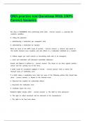
Cone & Rod receptors: absorb the light and first step of process of stimulus. In the receptors an electrical signal
is generated passed on to the bipolar cells, which passes it to the retinal ganglion cells that pass it on to the
optic nerve that passes it on to the brain.
Light comes in the other side, and hits the back of the eyeball, that how the process starts.
Horizontal cells and amacrine cells modulate the firing of the bipolar cells.
Inhibition happens in the horizontal cells.
Question about this on the exam: rod & Cone receptors density is not the same all across the retina, they have
different density across the retina. Cone density is the highest in the fovea, the peak density of rods is about 20
degrees of the fovea.
5-6 million cones
100-120 million rods
1 million ganglion cells.
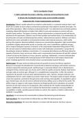
Thus, amount of receptors are much higher than the ganglion cells: many receptors converge (connect) to 1
ganglion cell. The strength of the convergence is not the same for rod and cones, it’s much stronger for the rod
receptors than cones e.g. in the fovea, there are individual cone cells that pass on their info directly to 1
ganglion cell.
This has consequences on our vision e.g. vision acuity: how much detail we can see with the rode and cones.
Suppose I have a tiny flashlight and shine it on the retina and just a single receptor. If I were to do that on 1
individual cone receptor, it will activate and pass it on to the ganglion cell that passes it on to the brain. If I
move the the flashlight to another individual cone, this happens too. Depending on where I shine my light, the
brain can distinguish diff signals because they’re all individual. This isn’t the case on the multiple rod receptors
that are connected to 1 ganglion cell; thus info gets lost because the brain cannot differentiate where the light
comes from, therefore we cannot see as many details with the rods as we do with the cones. That’s why you
cant see details from the corner of your eyes; because the light bouncing of the objects won’t go to the fovea
but beside it where the rods are. So you can see fine details with cones, not the rods.
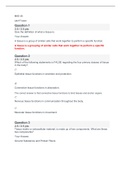
Consequences for light sensitivity (seeing in the dark). Individual cones: ganglion cell is lazy and wont activate if
the signal isn’t strong enough, no signal is passed on to the brain and so you won’t see anything. For the
multiple rods connected to 1 ganglion cell its diff, the signals of these rods get summated, which causes a signal
in ganglion cell which is passed on to the brain. This is why you can see better in the dark with the rods e.g. if
you’re outside on a clear night in the desert looking at a weak star, the moment you’re focusing on the star you
won’t be able to see it anymore because the light is projected on your fovea whose cones doesn’t’ send
messages to the brain because the light is too weak. If you move your eye to the left or right you’d see the star
again . You can see better in the dark with rod receptors.
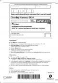
Lateral inhibition: sideways suppression. Which means that cells in the retina can cause activation of other cells
(e.g. bipolar cell can generate a signal in a ganglion cell), but they can also decrease the activation of other cell.
This plays a role in the perception of mach bands. Objectively the light intensity of the band is uniform, from left
to right it has the same intensity of light within each band. Yet, each band appears to be a bit darker on the left
than the right side. How? Caused by lateral inhibition. Horizontal cells play an important role e.g. receptors pass
on info to their cells, an arrow straight to other cells is an excitatory connection. With lateral inhibition
(negative input) that same cell passes info to the two cells on the left and right, but the strength of that
inhibition decreases.
Cone & Rod receptors: absorb the light and first step of process of stimulus. In the receptors an electrical signal
is generated passed on to the bipolar cells, which passes it to the retinal ganglion cells that pass it on to the
optic nerve that passes it on to the brain.
Light comes in the other side, and hits the back of the eyeball, that how the process starts.
Horizontal cells and amacrine cells modulate the firing of the bipolar cells.
Inhibition happens in the horizontal cells.
Question about this on the exam: rod & Cone receptors density is not the same all across the retina, they have
different density across the retina. Cone density is the highest in the fovea, the peak density of rods is about 20
degrees of the fovea.
5-6 million cones
100-120 million rods
1 million ganglion cells.
Thus, amount of receptors are much higher than the ganglion cells: many receptors converge (connect) to 1
ganglion cell. The strength of the convergence is not the same for rod and cones, it’s much stronger for the rod
receptors than cones e.g. in the fovea, there are individual cone cells that pass on their info directly to 1
ganglion cell.
This has consequences on our vision e.g. vision acuity: how much detail we can see with the rode and cones.
Suppose I have a tiny flashlight and shine it on the retina and just a single receptor. If I were to do that on 1
individual cone receptor, it will activate and pass it on to the ganglion cell that passes it on to the brain. If I
move the the flashlight to another individual cone, this happens too. Depending on where I shine my light, the
brain can distinguish diff signals because they’re all individual. This isn’t the case on the multiple rod receptors
that are connected to 1 ganglion cell; thus info gets lost because the brain cannot differentiate where the light
comes from, therefore we cannot see as many details with the rods as we do with the cones. That’s why you
cant see details from the corner of your eyes; because the light bouncing of the objects won’t go to the fovea
but beside it where the rods are. So you can see fine details with cones, not the rods.
Consequences for light sensitivity (seeing in the dark). Individual cones: ganglion cell is lazy and wont activate if
the signal isn’t strong enough, no signal is passed on to the brain and so you won’t see anything. For the
multiple rods connected to 1 ganglion cell its diff, the signals of these rods get summated, which causes a signal
in ganglion cell which is passed on to the brain. This is why you can see better in the dark with the rods e.g. if
you’re outside on a clear night in the desert looking at a weak star, the moment you’re focusing on the star you
won’t be able to see it anymore because the light is projected on your fovea whose cones doesn’t’ send
messages to the brain because the light is too weak. If you move your eye to the left or right you’d see the star
again . You can see better in the dark with rod receptors.
Lateral inhibition: sideways suppression. Which means that cells in the retina can cause activation of other cells
(e.g. bipolar cell can generate a signal in a ganglion cell), but they can also decrease the activation of other cell.
This plays a role in the perception of mach bands. Objectively the light intensity of the band is uniform, from left
to right it has the same intensity of light within each band. Yet, each band appears to be a bit darker on the left
than the right side. How? Caused by lateral inhibition. Horizontal cells play an important role e.g. receptors pass
on info to their cells, an arrow straight to other cells is an excitatory connection. With lateral inhibition
(negative input) that same cell passes info to the two cells on the left and right, but the strength of that
inhibition decreases.