Questions
Discuss the molecular diversity of ion channels
Different ion channels have different properties. One way to distinguish them is to look at their
selectivity for different ions. The more important ions for cell physiology are K+, Cl-, Na+ and
Ca2+ ions. Different channels will sometimes only be able to let through 1 ion type.
Related to this, the different channels will open at different potentials of the cell membrane
related to the reversal potential of this ion. These are voltage dependent channels. We can
calculate this portential using the Nernst eqation. (= 2.3 RT/zF * log ([X]o/[X]i)) The different
kinetics of an ion channel are also determined by the probability the channel is open at a given
timepoint.
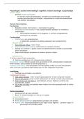
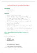
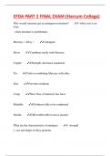
Another aspect is the I/V relationship which defines permeation. We can illustrate this with a
graph. Each type of ion channel can have a different conductance or rectification properties.
Rectification is the process of restabilizing the cell to rest potential. The channel (or other
proteins) could be seen as the resistor in a RC circuit with the cell membrane (the bilipid layer)
as the capacitor. Ohm’s law is then relevant here. (R=V/I
Lastly there can be a difference in effects on the channel from frugs, pharmaceuticals and toxins,
which can modify the behaviours of the channels.
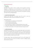
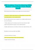
, Describe the crystal structure of K-channels in relation to the function
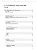
The kristals have a tetrameric structure with on the outside they are arrange around a central
hole which is the permeation pathway. You have 2 transmembranic alfa helices with a p-loop
in between per subunit. The transmembranic sheets are 20° turned away from being
permendicular to the membrane. In the kristal structure they can put in different ions and test
the model to be sure it is a K+.
1. tetramer
2. aromatic resdues at water lipid interface
3. Per subunit 2 alfa helices with a p-loop inbetween (p-loop goes only a little bit in)
4. Outerhelix- Turret/P – Helix/select filter – inner helix
5. Main helices at a 25° tilt + inner helix kink
6. Cone/tipi vorm, base + extracellular face
7. Base holds p region/ selectivity filter
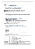
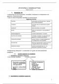
8. Pore helix: filling up withstructure of the p-loop and then makes a helix pointed to the
central cavity. Negativity of the dipole points towards the narrow part. (broad base)
a. Between inner and outer helices
b. Electronegative C-term towards the pore
c. Intersubunit contacts
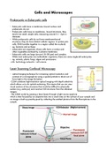
9. Pore:
a. 45 A long
b. Inside is 18 A long, 6 wide Hydrofobic lining
c. Central cavity is 10 A diameter Hydrofobic lining
d. Selectivity filter is12 A long Carbonyl oxygens
e. Two K+ binding sides: 7.5 A separated
f. (third side in cavity)
10. Function of the central cavity: lower electrostatic barrier for permeating cation
a. Hydrohylic environment
b. C-terminal end pore helix: electronegative
c. Note: no deep energy wells, binding in QA derivatives (TEA), reduction of
relevant diffusion distance to 12A
11. Selectivity filter:
a. Side chain GYG away from pore
b. 1 water molecular between 2 K ions
c. Main chain carbonyl oxygen = coordinate dehydrated K
d. Potassium has to dehydrate to get through the selectivity filter. Sadium can only
interact with 2 O ions so it prefers to stay with the 4 H molecules and can not
enter.
12. Packing around GYG: aromatics (12 in total)
a. Solid sheet
b. Hold GYG at correct distance for K but not Na
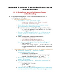
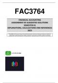
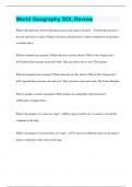
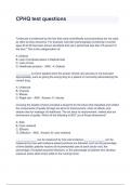
K-selectivity: 4 ~ cubical sites in selectivity filter (backbone oxygens)
Potassium ions positions in selectivity filter. Single file, Electrostatic repulsion. There are 4
sites that are important in the shaping of the channel depiced here. And the K+ can be briefly
in each box. Potassium ion in solution (depicted with 8H molecules in the hydration shell
There are at least 12 AZ devided over the 4 subunits that all interact with each other. The GYG
opens exactly enough to have an interaction with the K+ ions.
Discuss the molecular diversity of ion channels
Different ion channels have different properties. One way to distinguish them is to look at their
selectivity for different ions. The more important ions for cell physiology are K+, Cl-, Na+ and
Ca2+ ions. Different channels will sometimes only be able to let through 1 ion type.
Related to this, the different channels will open at different potentials of the cell membrane
related to the reversal potential of this ion. These are voltage dependent channels. We can
calculate this portential using the Nernst eqation. (= 2.3 RT/zF * log ([X]o/[X]i)) The different
kinetics of an ion channel are also determined by the probability the channel is open at a given
timepoint.
Another aspect is the I/V relationship which defines permeation. We can illustrate this with a
graph. Each type of ion channel can have a different conductance or rectification properties.
Rectification is the process of restabilizing the cell to rest potential. The channel (or other
proteins) could be seen as the resistor in a RC circuit with the cell membrane (the bilipid layer)
as the capacitor. Ohm’s law is then relevant here. (R=V/I
Lastly there can be a difference in effects on the channel from frugs, pharmaceuticals and toxins,
which can modify the behaviours of the channels.
, Describe the crystal structure of K-channels in relation to the function
The kristals have a tetrameric structure with on the outside they are arrange around a central
hole which is the permeation pathway. You have 2 transmembranic alfa helices with a p-loop
in between per subunit. The transmembranic sheets are 20° turned away from being
permendicular to the membrane. In the kristal structure they can put in different ions and test
the model to be sure it is a K+.
1. tetramer
2. aromatic resdues at water lipid interface
3. Per subunit 2 alfa helices with a p-loop inbetween (p-loop goes only a little bit in)
4. Outerhelix- Turret/P – Helix/select filter – inner helix
5. Main helices at a 25° tilt + inner helix kink
6. Cone/tipi vorm, base + extracellular face
7. Base holds p region/ selectivity filter
8. Pore helix: filling up withstructure of the p-loop and then makes a helix pointed to the
central cavity. Negativity of the dipole points towards the narrow part. (broad base)
a. Between inner and outer helices
b. Electronegative C-term towards the pore
c. Intersubunit contacts
9. Pore:
a. 45 A long
b. Inside is 18 A long, 6 wide Hydrofobic lining
c. Central cavity is 10 A diameter Hydrofobic lining
d. Selectivity filter is12 A long Carbonyl oxygens
e. Two K+ binding sides: 7.5 A separated
f. (third side in cavity)
10. Function of the central cavity: lower electrostatic barrier for permeating cation
a. Hydrohylic environment
b. C-terminal end pore helix: electronegative
c. Note: no deep energy wells, binding in QA derivatives (TEA), reduction of
relevant diffusion distance to 12A
11. Selectivity filter:
a. Side chain GYG away from pore
b. 1 water molecular between 2 K ions
c. Main chain carbonyl oxygen = coordinate dehydrated K
d. Potassium has to dehydrate to get through the selectivity filter. Sadium can only
interact with 2 O ions so it prefers to stay with the 4 H molecules and can not
enter.
12. Packing around GYG: aromatics (12 in total)
a. Solid sheet
b. Hold GYG at correct distance for K but not Na
K-selectivity: 4 ~ cubical sites in selectivity filter (backbone oxygens)
Potassium ions positions in selectivity filter. Single file, Electrostatic repulsion. There are 4
sites that are important in the shaping of the channel depiced here. And the K+ can be briefly
in each box. Potassium ion in solution (depicted with 8H molecules in the hydration shell
There are at least 12 AZ devided over the 4 subunits that all interact with each other. The GYG
opens exactly enough to have an interaction with the K+ ions.