Samenvatting MRI
Lecture 1
- What is needed for MR imaging
- Nature of the MR signal
- Image contrast
Information
- Animal model: for a specific disease, we try to initiate an animal model
o Knock-out, knock-in, …
o Allows us to study the disease by itself in a quite fast way
o To have an early biomerker of the disease
o Big advantages of MRI:
▪ All have the same modality, but will have different contrasts
▪ Doesn’t allow only 1 image, but you can change the contrast
• You can specify it as an user -> to get the optimal contrast
Different modalities that can be used to do in Vivo imaging
- Most will use EM-waves:
o X-ray
▪ CT and RX
o Radiowaves
▪ MRI/spectroscopy
o Gammawave (nucleair medicine)
▪ PET (positron emission tomography)
▪ SPECT (single photon emission computed tomography)
- Without EM-waves:
o Ultrasounds
Spectrum of EM-waves
- Have a certain frequency, wavelength and
energy
- Photon of EM-wave of a certain frequency f
is actually linearity related to the energy of
the photons
o Higher frequency -> higher energy
content of the EM-waves
o Higher number of photons -> higher
the intensity of wave
- Radiowaves:
o Used for MRI
o Very small energy -> not dangerous
o MRI can be performed repeatedly (for humans)
1
, - X-ray and gamma-ray
o Radioprotection!
o To protect the patient, nurse, family and doctor
o Causing possible harm to DNA and might induce cancer
Introduction
- Basic signal for the most MRI-imaging comes from the hydrogen-nucleus (proton)
o Hydrogen-nucleus belongs to a water molecule
▪ = substance that will give the signal intensity in the end
o Contrast -> proton density
▪ Higher amount of protons as compared to another tissues -> signal
is related to that proton density
▪ Can create that contrast
- How protons interacted with the surrounding tissue (biochemical environment)
will contribute to MRI specific parameters
o Parameters: only have a meaning in the MRI -> related to the relaxation
times (T1, T2, T2*)
▪ Can be dependent to the biochemical environment
▪ Can have the same proton density in different tissues, but when
they have a different T1 -> T1 weighted image will show you
differences between these 2 tissues
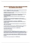
- Images: T2-weighted images
o T2: relaxation time related to the water content
▪ Higher water content -> higher T2 value
▪ Ventricles (brain) filled with CSF = very bright
o Results:
▪ Enlarged ventricle volumes
• Volume reduction of the entire brain
• Abnormal shape of the cerebellum
o Look at the respons for certain drugs
- Image of MRI:
o 2D
▪ Image will be divided into pixels
▪ Pixels = picture element
▪ Each pixel has a certain value -> matrix
• Will specific the intensity in your image
▪ Will give you a certain intensity or grey level
o 3D: Taking a slice with a certain thickness
▪ Same grid as your image
▪ The signal intensity comes from a volume element
▪ Slice is divided in subparts
2
, • Voxelelements is filled with your watermolecule
• Protons are the source of the signal
▪ Look at the property of the protons in dedicated voxel
▪ Dependent on which contrast we want we can change if that
property will give a bright or dark signal intensity
▪ Which property you are going to visualize? -> this you can change
o Image = population of protons in a small voxel with a certain slice
thickness
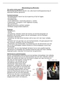
Different image contrast
- All acquired with the same sequence
- Only 2 time parameters are changed
- Get a complete different image
o Complete different contrast
- Different images:
o T1-weighted: making contrast
depending on T1 differences
o T2-weighted: differences in the
T2
Other modalities
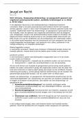
- MRI also allows you to capture another contrast, like:
o Diffusion
▪ Diffusion-MRI: follows the random motion of the
water molecules
▪ Watermolecules diffusion in human brain are
confronted with restrictions
• Will hit the external membranes
• Restricted by myeline
• Only move in a certain directions
▪ Get information about the white matter
▪ Colour specifies the direction
▪ Measure demyelinisation
o Tissue perfusion
▪ Cerebral blood flow
o Blood volume
▪ In the major blood vessels
o Angiograms
▪ Images of the blood vessels
▪ With/without contrast agent
o Activated brain regions = functional MRI (fMRI)
▪ Which regions in the brain is activated with you do a specific task
3
, ▪ In animals: electrical stimulation
Information provided
- Different informationtypes:
o Anatomical information
o Physiological information
o Functional information
o Molecular information, migration of labeled stem cellls
- Few other nuclei may be used for specialized imaging purposes
- MRI: particularly suited to imaging differences between soft tissues, such as in
the head, neck and spiral regions of the body
Synopsis of MRI
- = short synopsis of MRI
- Need a huge magnet = basic compound of the MRI-scanner
o Big coil
o Need to generate a very high magnetic field
o Current flow going through that coil
o Right hand rule
o Very high magnetic field in the inner side -> very big coil through which
very high current is flowing
- Field strengths = very high
o Most hospitals: 3T
o MRI on small animals: size of volume elements are quite different
▪ Lose in volume -> go 1000 times smaller
▪ Volumes from which go capture your signal will be 1000 times
smaller -> need more signal intensity
▪ Higher field strength -> increase the signal you will capture
▪ 11,5T
- Magnets are superconducting magnets
o Superconduct: no resistance
▪ Resistance is dependent on the temperature
▪ Cool down the coil the resistance will decrease
• Up to temperature of 4K -> by immersion the coil in liquid
helium -> there wille be no longer a resistance
▪ Install the scanner: 2 plugs -> put a high potential over those 2
cables -> increasing the current -> increasing the magnetic field
until the magnetic field is 11,5 T
• Stop and remove the plugs
• Scanner by itself is nolonger connected to electricity
• Current is always going on = dangerous part
4
Lecture 1
- What is needed for MR imaging
- Nature of the MR signal
- Image contrast
Information
- Animal model: for a specific disease, we try to initiate an animal model
o Knock-out, knock-in, …
o Allows us to study the disease by itself in a quite fast way
o To have an early biomerker of the disease
o Big advantages of MRI:
▪ All have the same modality, but will have different contrasts
▪ Doesn’t allow only 1 image, but you can change the contrast
• You can specify it as an user -> to get the optimal contrast
Different modalities that can be used to do in Vivo imaging
- Most will use EM-waves:
o X-ray
▪ CT and RX
o Radiowaves
▪ MRI/spectroscopy
o Gammawave (nucleair medicine)
▪ PET (positron emission tomography)
▪ SPECT (single photon emission computed tomography)
- Without EM-waves:
o Ultrasounds
Spectrum of EM-waves
- Have a certain frequency, wavelength and
energy
- Photon of EM-wave of a certain frequency f
is actually linearity related to the energy of
the photons
o Higher frequency -> higher energy
content of the EM-waves
o Higher number of photons -> higher
the intensity of wave
- Radiowaves:
o Used for MRI
o Very small energy -> not dangerous
o MRI can be performed repeatedly (for humans)
1
, - X-ray and gamma-ray
o Radioprotection!
o To protect the patient, nurse, family and doctor
o Causing possible harm to DNA and might induce cancer
Introduction
- Basic signal for the most MRI-imaging comes from the hydrogen-nucleus (proton)
o Hydrogen-nucleus belongs to a water molecule
▪ = substance that will give the signal intensity in the end
o Contrast -> proton density
▪ Higher amount of protons as compared to another tissues -> signal
is related to that proton density
▪ Can create that contrast
- How protons interacted with the surrounding tissue (biochemical environment)
will contribute to MRI specific parameters
o Parameters: only have a meaning in the MRI -> related to the relaxation
times (T1, T2, T2*)
▪ Can be dependent to the biochemical environment
▪ Can have the same proton density in different tissues, but when
they have a different T1 -> T1 weighted image will show you
differences between these 2 tissues
- Images: T2-weighted images
o T2: relaxation time related to the water content
▪ Higher water content -> higher T2 value
▪ Ventricles (brain) filled with CSF = very bright
o Results:
▪ Enlarged ventricle volumes
• Volume reduction of the entire brain
• Abnormal shape of the cerebellum
o Look at the respons for certain drugs
- Image of MRI:
o 2D
▪ Image will be divided into pixels
▪ Pixels = picture element
▪ Each pixel has a certain value -> matrix
• Will specific the intensity in your image
▪ Will give you a certain intensity or grey level
o 3D: Taking a slice with a certain thickness
▪ Same grid as your image
▪ The signal intensity comes from a volume element
▪ Slice is divided in subparts
2
, • Voxelelements is filled with your watermolecule
• Protons are the source of the signal
▪ Look at the property of the protons in dedicated voxel
▪ Dependent on which contrast we want we can change if that
property will give a bright or dark signal intensity
▪ Which property you are going to visualize? -> this you can change
o Image = population of protons in a small voxel with a certain slice
thickness
Different image contrast
- All acquired with the same sequence
- Only 2 time parameters are changed
- Get a complete different image
o Complete different contrast
- Different images:
o T1-weighted: making contrast
depending on T1 differences
o T2-weighted: differences in the
T2
Other modalities
- MRI also allows you to capture another contrast, like:
o Diffusion
▪ Diffusion-MRI: follows the random motion of the
water molecules
▪ Watermolecules diffusion in human brain are
confronted with restrictions
• Will hit the external membranes
• Restricted by myeline
• Only move in a certain directions
▪ Get information about the white matter
▪ Colour specifies the direction
▪ Measure demyelinisation
o Tissue perfusion
▪ Cerebral blood flow
o Blood volume
▪ In the major blood vessels
o Angiograms
▪ Images of the blood vessels
▪ With/without contrast agent
o Activated brain regions = functional MRI (fMRI)
▪ Which regions in the brain is activated with you do a specific task
3
, ▪ In animals: electrical stimulation
Information provided
- Different informationtypes:
o Anatomical information
o Physiological information
o Functional information
o Molecular information, migration of labeled stem cellls
- Few other nuclei may be used for specialized imaging purposes
- MRI: particularly suited to imaging differences between soft tissues, such as in
the head, neck and spiral regions of the body
Synopsis of MRI
- = short synopsis of MRI
- Need a huge magnet = basic compound of the MRI-scanner
o Big coil
o Need to generate a very high magnetic field
o Current flow going through that coil
o Right hand rule
o Very high magnetic field in the inner side -> very big coil through which
very high current is flowing
- Field strengths = very high
o Most hospitals: 3T
o MRI on small animals: size of volume elements are quite different
▪ Lose in volume -> go 1000 times smaller
▪ Volumes from which go capture your signal will be 1000 times
smaller -> need more signal intensity
▪ Higher field strength -> increase the signal you will capture
▪ 11,5T
- Magnets are superconducting magnets
o Superconduct: no resistance
▪ Resistance is dependent on the temperature
▪ Cool down the coil the resistance will decrease
• Up to temperature of 4K -> by immersion the coil in liquid
helium -> there wille be no longer a resistance
▪ Install the scanner: 2 plugs -> put a high potential over those 2
cables -> increasing the current -> increasing the magnetic field
until the magnetic field is 11,5 T
• Stop and remove the plugs
• Scanner by itself is nolonger connected to electricity
• Current is always going on = dangerous part
4