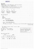
3 - COLUMNS AND PATHWAYS
RECAP
- Iris: regulates the amount of light entering the eyeball
- Cornea, lens & vitreous humors: focus light rays so that clear image is formed on the retina
- Rod & cones receptors: capture that image
- Postreceptoral layers: translate the raw light array captured by the photoreceptors into patterns of
spot surrounded by darkness, or vice versa
- Ganglion cells: detected all that
- Retinal translation: helps us perceive the pattern of light and dark areas in the visual field,
regardless of the overall light level
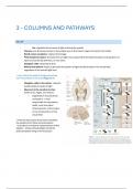
→ now: follow the path of image processing
from the eyeball to the brain (Figure 3.1)
- Ganglion cells in the retina: respond
preferentially to spots of light
- Neurons in the cerebral cortex:
prefer lines, edges, and stripes
o Organized in thousand tiny
computers -> each
responsible for orientation,
width, color and other
characteristics of the stripes
in one small portion of the
visual field
→ How do other parts of the brain assemble
the outputs from these minicomputers
- to produce a coherent representation of the
objects - whose reflected light stared the
photoreceptors firing in the first place
,4.1 FROM RETINA TO VISUAL CORTEX
How does the visual signal get from the retina to the visual area of the cortex?
PATHWAY TO THE BRAIN
• Visual signals from both eyes leave the back of the eye in the optic nerve and meet at the optic
chiasm à x-shaped bundle of fibers on the ventral
surface of the brain, inferior to the hypothalamus
• At the optic chiasm, some of the fibers cross to the
contralateral brain hemisphere
o All fibers corresponding to the right visual field
(not eye) end up on the left hemisphere and vice
versa. Each hemisphere processes the input
from the ipsilateral side of each retina, which
corresponds to the contralateral visual field
o The visual field is determined based on the point
of fixation:
§ The left visual field is anything to the left
of the fixation and that is processed by the right hemisphere
§ The right visual field is anything to the right of the fixation point and that is
processed by the left hemisphere
AFTER THE OPTIC CHIASM CROSSOVER
• 90% of the signals from the retina proceed to the Lateral Geniculate Nucleus (LGN) in the thalamus
of each hemisphere
• 10% of fibers travel to the Superior Colliculus (involved in controlling eye movements)
• Thalamus serves as a relay station where incoming sensory information makes a stop before
reaching the cerebral cortex
LGN – LATERAL GENICULATE
NUCLEUS
• Neurons in the LGN also have
center-surround receptive fields
• The signal sent from the LGN to the
cortex is smaller than the input the
LGN receives from the retina
• LGN regulates neural information as
it flows from the retina to the cortex
, • NOT ALL INFO GOES TO HIGHER ORDER AREAS; more feedback back, bc relay station
• LGN also takes info from cortex back and decides what goes to higher order
• Thalamus Integrates info from all senses and decides what needs more processing
1.
ð left LGN receives projections from the left side of the retina in both eyes ?
ð right LGN receives projections from the right side of both retinas ?
2.
ð Each layer of the LGN receives input from one or the other eye
From bottom to top
- Layers 1,4 and 6 of the right LGN receive input from the left eye (contralateral)
- Layer 2,3, and 5 get their input from the right eye (ipsilateral)
- Each LGN layer contains a highly organized map of a complete half of the visual
field = topographical mapping = orderly mapping of the world in the lateral
geniculate nucleus and the visual cortex
The axons of retinal ganglion cells synapse in the two lateral geniculate nuclei (LGNs),
on each hemisphere
ð these act as relay stations from the retina to the cortex
ð six-layered structure
ð the neurons in the bottom two layers are physically larger than those in the top four layers
o bottom two = magnocellular layers
o top four = parvocellular layers
magnocellular layers
parvocellular layers
Bottom two (1-2) Top four (3-6)
Receive input from M ganglion cells Receive input from P ganglion cells
Respond two large, fast-moving objects Responsible for processing details of stationary
targets
ð visual system splits input from the image into different types of information
ð between the magno & parvo layers = koniocellular cells => each koniocellular layer seems to be
involved in a different aspect of processing
o e.g., one is specialized for relaying signals from the S-cones and may be part of a “primordial” blue-
yellow
RECAP
- Iris: regulates the amount of light entering the eyeball
- Cornea, lens & vitreous humors: focus light rays so that clear image is formed on the retina
- Rod & cones receptors: capture that image
- Postreceptoral layers: translate the raw light array captured by the photoreceptors into patterns of
spot surrounded by darkness, or vice versa
- Ganglion cells: detected all that
- Retinal translation: helps us perceive the pattern of light and dark areas in the visual field,
regardless of the overall light level
→ now: follow the path of image processing
from the eyeball to the brain (Figure 3.1)
- Ganglion cells in the retina: respond
preferentially to spots of light
- Neurons in the cerebral cortex:
prefer lines, edges, and stripes
o Organized in thousand tiny
computers -> each
responsible for orientation,
width, color and other
characteristics of the stripes
in one small portion of the
visual field
→ How do other parts of the brain assemble
the outputs from these minicomputers
- to produce a coherent representation of the
objects - whose reflected light stared the
photoreceptors firing in the first place
,4.1 FROM RETINA TO VISUAL CORTEX
How does the visual signal get from the retina to the visual area of the cortex?
PATHWAY TO THE BRAIN
• Visual signals from both eyes leave the back of the eye in the optic nerve and meet at the optic
chiasm à x-shaped bundle of fibers on the ventral
surface of the brain, inferior to the hypothalamus
• At the optic chiasm, some of the fibers cross to the
contralateral brain hemisphere
o All fibers corresponding to the right visual field
(not eye) end up on the left hemisphere and vice
versa. Each hemisphere processes the input
from the ipsilateral side of each retina, which
corresponds to the contralateral visual field
o The visual field is determined based on the point
of fixation:
§ The left visual field is anything to the left
of the fixation and that is processed by the right hemisphere
§ The right visual field is anything to the right of the fixation point and that is
processed by the left hemisphere
AFTER THE OPTIC CHIASM CROSSOVER
• 90% of the signals from the retina proceed to the Lateral Geniculate Nucleus (LGN) in the thalamus
of each hemisphere
• 10% of fibers travel to the Superior Colliculus (involved in controlling eye movements)
• Thalamus serves as a relay station where incoming sensory information makes a stop before
reaching the cerebral cortex
LGN – LATERAL GENICULATE
NUCLEUS
• Neurons in the LGN also have
center-surround receptive fields
• The signal sent from the LGN to the
cortex is smaller than the input the
LGN receives from the retina
• LGN regulates neural information as
it flows from the retina to the cortex
, • NOT ALL INFO GOES TO HIGHER ORDER AREAS; more feedback back, bc relay station
• LGN also takes info from cortex back and decides what goes to higher order
• Thalamus Integrates info from all senses and decides what needs more processing
1.
ð left LGN receives projections from the left side of the retina in both eyes ?
ð right LGN receives projections from the right side of both retinas ?
2.
ð Each layer of the LGN receives input from one or the other eye
From bottom to top
- Layers 1,4 and 6 of the right LGN receive input from the left eye (contralateral)
- Layer 2,3, and 5 get their input from the right eye (ipsilateral)
- Each LGN layer contains a highly organized map of a complete half of the visual
field = topographical mapping = orderly mapping of the world in the lateral
geniculate nucleus and the visual cortex
The axons of retinal ganglion cells synapse in the two lateral geniculate nuclei (LGNs),
on each hemisphere
ð these act as relay stations from the retina to the cortex
ð six-layered structure
ð the neurons in the bottom two layers are physically larger than those in the top four layers
o bottom two = magnocellular layers
o top four = parvocellular layers
magnocellular layers
parvocellular layers
Bottom two (1-2) Top four (3-6)
Receive input from M ganglion cells Receive input from P ganglion cells
Respond two large, fast-moving objects Responsible for processing details of stationary
targets
ð visual system splits input from the image into different types of information
ð between the magno & parvo layers = koniocellular cells => each koniocellular layer seems to be
involved in a different aspect of processing
o e.g., one is specialized for relaying signals from the S-cones and may be part of a “primordial” blue-
yellow