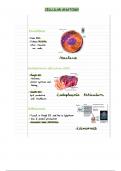
HLSC120 – Human Anatomy
Chapter 6: Bones
Skeleton – supporting framework for the soft tissues of the body Interstitial growth – declines rapidly as the cartilage matures bec the matrix is
Skeletal system – composed of dynamic living tissues; interacts w/ all of the other no longer able to expand
organ systems; continually rebuilds and remodels itself; includes bones, cartilage, 1. chondrocytes w/n the lacunae divide to form 2 chondroblasts
ligaments, connective tissues that stabilize or connect the bones 2. cells grow, begin to produce new matrix and push apart from each
CARTILAGE other forming 2 new chondrocytes
semi-rigid, connective tissue found throughout the human body; weaker than bone, Appositional growth – where further growth occurs later on
but more flexible and resilient; contains cells that are scattered throughout an 1. cartilage grows through the division of stem cells at the internal edge
abundant matrix of protein fibers embedded w/n a gel-like ground substance; of the perichondrium
avascular 2. differentiation of committed cells into chondroblasts results in the
Chondroblasts – cells that produce the matrix of cartilage formation of new cartilage matrix and the differentiation of these cells
Chondrocytes – cartilage cells; secreted by the chondroblasts into chondrocytes w/n the inner layer of the perichondrium
Lacunae – small spaces occupied by the chondrocytes BONE
Functions: support our weight and interact with muscles to produce precisely controlled
Support soft tissues – C- shaped hyaline cartilage rings for trachea support; movements; complex, dynamic organs composed of all tissue types:
flexible cartilage supports the auricle of the ear Bone connective tissue – osseous connective tissue; primary component;
Provides a gliding surface at articulations/joints where 2 bones meet matrix is sturdy and rigid due to calcification
Provides a model for the formation of most of the bones in the body; cartilage Calcification – mineralization; deposition of minerals in the matrix
serves as a rough draft that is later replaced by bone tissue Connective tissue proper – periosteum
Types of Cartilage: Cartilage connective tissue – articular cartilage
Hyaline – most abundant; found in trachea, larynx, articular (joint) cartilage on Smooth muscle tissue – forming the walls of blood vessels that supply bone
bones, epiphyseal plates, and the fetal skeleton; provides support through Fluid connective tissue – blood
flexibility and resilience; translucent appearance; surrounded by perichondrium Epithelial tissue – lining of the blood vessels
Perichondrium – dense connective tissue covering of hyaline cartilage Nervous tissue – nerves that supply bone
Fibrocartilage – has extracellular matrix w/ numerous thick collagen fibers that Functions:
help resist both tensile (stretching) and compressional (compaction) forces; can Support and protection
act as a shock absorber; located in intervertebral discs, menisci of the knee, Movement
pubic symphysis; lacks a perichondrium bec stress applied at the surface of the Hemopoiesis – process of blood cell production
fibrocartilage would quickly destroy this later Red bone marrow – a connective tissue where blood cells are
Elastic – contains elastin w/n its extracellular matrix; found in auricle of the produced; located in some spongy bone; contains stem cells that form
external ear, external auditory canal, epiglottis all of the blood cell types; found in adults’ flat bones of the skull,
Elastin – highly branched elastic fibers vertebrae, ribs, sternum (breastbone), ossa coxae (hip bones),
Growth patterns of cartilage – once the cartilage is fully mature, new cartilage proximal epiphyses of the humerus and femur
growth typically stops entirely, from this point on, cartilage growht usually occurs Yellow bone marrow – when the red bone marrow degenerates and
only after injury to the cartilage turns into a fatty tissue; located in shafts of long bones
Storage of mineral and energy reserves
, Calcium – essential for muscle contraction, blood clotting and nerve o periosteum – tough sheath; covers the outer surface of the bone w/
impulse transmission the exception of areas covered by articular cartilagee; made of dense
Phosphate – needed for ATP utilization irregular connective tissue; consists of an outer fibrous layer and an
Classification of Bones: inner cellular layer; isolates and protects the bone from surrounding
Long bones – have greater length than width; elongated, cylindrical shaft structures, anchors blood vessels and nerves to the surface of the
(diaphysis) has distinct ends (epiphyses); found in the upper limb (arm, bone; provides stem cells (osteoprogenitor cells and osteoblasts) for
forearm, palm, fingers) and longer limb (thigh, leg, sole of the foot, toes); most bone width growth and fracture repair; anchored to the bone by
common type; serve as a useful model of bone structure; perforating fibers
2 types: perforating fibers – tough collagen fibers; run perpendicular
o femur – thigh bone to the bone shaft
o humerus – arm bone Short bones – have a length nearly equal to their width; external surfaces are
Parts: covered by compact bone and interior are covered by spongy bone; examples
o diaphysis – principal gross feature; shaft; elongated, usually cylindrical; are carpals (wrist bones) and tarsals (bones in the foot), sesamoid bones (tiny,
provides for the leverage of a long bone seed-shaped bones along the tendons of some muscles), patella (kneecap,
medullary cavity – hollow, cylindrical space w/n the diaphysis; largest sesamoid bone)
in adults, contains yellow bone marrow; often referred to as Flat bones – have a flat, thin surface; composed of roughly parallel surfaces of
marrow cavity compact bone w/ a layer of internally placed spongy bone; provide extensive
o epiphysis – located at each end of along bone; expanded, knobby surfaces for muscle attachment; protect underlying soft tissues; form the roof
region; enlarged to strengthen the joint; provide added surface area of the skull, scapulae (shoulder blades); sternum (breastbone), ribs
for tendon and ligament attachment; composed of an outer layer of Irregular bones – have elaborate, complex shapes; vertebrae and several bones
compact bone and inner layer of spongy bone in the skull (ethmoid and sphenoid bones) are examples of it
proximal epiphysis – end of the bone closest to the body Cells of bone:
trunk Osteoprogenitor cells – stem cells derived from mesenchyme; when they
distal epiphysis – end farthest from the trunk divide, they produce another stem cell and a committed cell that matures to
o metaphysis – region in a mature bone sandwiched b/w the diaphysis become an osteoblast; located w/n both the periosteum and endosteum
and the epiphysis Osteoblasts – formed from osteoprogenitor cells; exhibit a cuboidal structure;
epiphyseal (growth) plate – contained in the metaphysis; thin secrete osteoid; produce new bone; when entrapped in the matrix, they
layers of hyalne cartilage that provide for the continued produce and secrete and differentiate into osteocytes
lengthwise growth of the diaphysis Osteoid – initial semisolid form of bone matrix; calcifies as a result of
epiphyseal line – remnant of the epiphyseal plate; thin, calcium deposition
defined area of the compact bone Osteocytes – mature bone cells derived from osteoblasts that have become
o articular cartilage – thin layer of hyaline cartilage covering the entrapped in the matrix they secreted; reside in small spaces w/n the matrix
epiphysis at a join surface; helps reduce friction, absorb shock in (lacunae); maintain the bone matrix; detect mechanical stress on a bone; this
moveable joints info is communicated to osteoblasts and may result in the deposition of new
o endosteum – an incomplete cellular membrane that covers all internal bone matrix at the surface
surfaces of the bone such as the medullary cavity; contains Osteoclasts – large, multinuclear, phagocytic cells; derived from bone marrow
osteoprogenitor cells, osteoblasts, and osteoclasts; active during bone cells; exhibit a ruffled border where they contact the bone w/c increases their
growth, repair, and remodeling surface area exposure to the bone; located w/n or adjacent to