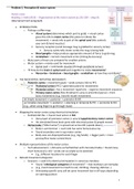
Problem 1: Perception & motor system:
Part 0: Intro:
Reading 1: Kolb (2015) – Organization of the motor system (p.232-250 – chap.9):
(Skip Spinal Cord paragraph)
• INTRODUCTION:
o Picking a coffee mug:
Visual system (determines which part to grab) -> visual cortex
gives this info to motor cortex (this plans to initiate the
movement) -> sends info to part of the spinal cord (this controls
your arm & hand muscles)
Sensory receptors (send message mug is grabbed to sensory cortex)
• Sensory cortex tells motor cortex the mug is being held
Basal ganglia = helps produce appropriate amount of force to grab mug
Cerebellum = corrects movement errors (timing & accuracy)
o Many parts of brain are activated for smallest actions
o Motor system = mainly used for movement
Spinal cord -> commands muscles (through peripheral nerves)
o How do the brain & spinal cord work together to make movements?
Neocortex + brainstem + basal ganglia + cerebellum how they contribute?
• THE NEOCORTEX: INITIATING MOVEMENT:
o Posterior cortex = movement goals – sends sensory info to PFC
Prefrontal cortex (PFC) = generates plans for movements
Premotor cortex = has a movement repertoire – organize movement sequence
Primary motor cortex (M1; Brodmann’s area 4; precentral gyrus) = more
basic movements (e.g., hand & mouth movement)
Simple movement => posterior -> premotor & M1
Complex movement => posterior -> planning in temporal & PFC -> premotor & M1
(e.g., when using finger to go through maze)
• Mapping the motor cortex using electrical stimulation:
o Penfield did this -> found most action in M1
Dorsal part of premotor cortex (= area 6/supplementary motor cortex)
He stimulated these brain parts with electrical pulses & people moved
o Homunculus (“little human”) is spread out across M1 (upside down)
Body is symmetrical -> each side has same homunculus rep
Found secondary one in sup motor cortex
Body sizes are disproportionate (e.g., big hands) -> bigger parts = more
precise/fine motor control there
• Multiple representations of the motor cortex:
o Tech advancement -> did same as Penfield but with microelectrodes = found many
more homunculi (≈10) – probs not as simple as he sketched
E.g., different locations for each finger
• Natural movement categories:
o Graziano -> used 0.5s electrical stimulation in conscious monkeys
Found “ethological categories of movement” – that monkeys use everyday
(e.g., hand movement to mouth to eat; climbing; chewing; defense…)
Stimulating specific area will do same movement but in dif ways depending
on starting position
1
, Hand stays there if keep stimulating + :/ hits object if it’s in the way
o 3 types of organization in each region (body part to move; special location of where
movement is directed; movement function)
Flexible map (depends on past/recent experience, objects available)
o Penfield + Graziano = whole body movement in dorsal premotor cortex
Hand/reaching is ventrally
Hand movement to mouth in most ventral part of premotor cortex
-> Whole body movements more in premotor cortex + more discrete in M1
• Visual-parietal-motor connections:
o Can also get movements by stimulating parietal cortex
Parietal topography = mirrors homunculus
Stepping -> dorsal – reaching -> medial – hand & mouth -> ventral
o E.g., reaching = visual cortex (shape & location) -> parietal (id body part that will
contact the object-reach out) -> motor (moves arm to grab it)
Movement itself = from brainstem or spinal cord
Movement = premotor (whole body) + M1 (discrete) – but also the parietal cortex!!
- Homunculus (Penfield)
• The movement lexicon:
o Graziano -> support for movement lexicon
E.g., many mammals use pincer grip (thumb & index) to grab stuff -> lesions
in thumb area of homunculus = weak thumb + poor coordination linked to it
(i.e., can’t pincer grip)
-> This stuff ≠ just learned – but part of vocab in pre-wired lexicon
o Different in every mammal (more complex in humans)
o Premotor cortex repertoire = more complex > M1 (specific stuff)
E.g., monkey can make hand movements (M1) but not coordinate them (PM)
o Fukuda -> learning to move = learning how to use pre-organized movement
o Motor cortex -> plans action – firing more pre-lifting (+ more when heavy)
Movement direction
Monkey with lever -> move towards him = max neuron discharge
• The discharge reduces as you go more far away from this position
• Motor cortex = calculates direction + distance
• Mirroring movement:
o Activity in premotor cortex when making movement + when seeing someone else
do this movement (≈sympathy)
= Mirror system neurons (= encode goal of an action)
Doesn’t always happen (e.g., only f object within reach)
Some still respond when change in target/size
• Some can fill in the blanks (e.g., when only see part of movement)
o Monkeys -> core mirror neurons = more broadly tuned – wide range of actions for
obtaining a goal (= transitive movements) – includes parietal/motor circuit
o Humans -> core mirror neurons = transitive movements (= goal) – includes Broca’s area
Distributed system = respond to intransitive movements (≠ goal)
Flexible properties of mirror neurons = ability to imagine movements
• Control BCIs
o Mirror neuron theory = we understand own & other’s actions by internally
replicating the movement for it
-> Self/social-awareness + awareness of intention/action of others
Probably important for verbal & gestural language
Lack of empathy (seen in autism) -> maybe cause of mirror neuron dysfunction
2
, Mirror neurons = internally replicate other’s actions -> helps us understand their actions
- Helps us imagine movements / Part of premotor cells
Core mirror neurons = transitive movements (= goal) – in ventral parietal + ventral premotor
& motor
• THE BRAINSTEM: MOTOR CONTROL: (automatic movements)
o 26 pathways from brain stem to spinal cord
Info on balance & posture – control automatic nervous system
Often whole body movements (≠ neocortex)
o Hess -> implemented electrodes into brain of animals – then to
stimulating lead
Stimulate part = sudden movement (e.g., erect hair in cat)
Could also modulate animal’s emotional behavior (e.g., excitement vs fear)
o Brainstem functions = posture; standing up; coordinate movements; swimming;
walking; drinking; sex; grooming…
• The basal ganglia & movement force: (non-automatic movements)
o Basal ganglia = collection of subcortical nuclei in forebrain – connect motor
cortex to midbrain + sensory regions of neocortex to motor cortex
Caudate putamen (= large cluster of nuclei under frontal cortex)
• Part of it extends into a tail in temporal lobe – ending in amygdala
o BG = receives input from 2 main sources
All areas of neocortex & limbic cortex (includes motor) project to it
Nigrostriatal dopamine pathway (from substantia nigra)
• (Vice versa -> BG sends back to motor & substantia nigra)
Caudate + Putamen = main part of basal ganglia (part of striata)
- Basal ganglia = send & receive to motor (neocortex) + substantia nigra (force control)
o Movement disorders caused by damage in basal ganglia:
1. Damage in caudate putamen -> leads to unwanted movements
(dyskinesias – hyperkinetic)
• Seen in Huntington’s disease + Tourette’s syndrome
2. Cells of basal ganglia left intact + damaged input -> difficulty making
movements (hypokinetic symptoms) – loss of dopamine from sub nigra
• Parkinson’s disease (loss dopamine cells in substantia nigra + their
input into the basal ganglia)
=> So one of its major functions must be to modulate movement
Problems with basal ganglia = hypo/hyperkinetic / so must link to movement
o Keele & Ivry -> BG’s underlying function = generate force required per movement
Tested by asking people to press button (force determined line length)
• (People with BG dysfunction pressed to
hard/lightly
o Redgrave -> 2 pathways to motor cortex
Excitatory & inhibitory pathways
If indirect pathway dominates -> excitation of
GPi = inhibits thalamus = lower movement
If direct pathway -> inhibition in GPi = more
activity in thalamus = amplifies movement
Globus pallidus (GPi) = where both paths
converge – determines strength of movement
• Lower/destroying/stimulate it this can help Parkinson’s patients
Inhibitory/indirect = high GPi -> low thalamus -> low movement
3
, Excitatory/direct = low GPi -> high thalamus -> high movement
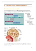
• The cerebellum & motor learning:
o Cerebellum = acquiring + maintaining motor skills
Under cerebral cortex – above brain stem
Smaller but contain 4x more neurons than neocortex
o Flocculus (small lobe on ventral surface of C – eye movement & balance)
Homunculus (middle linked to face…)
o Pathway between 2 hemispheres/midline -> cerebellar nuclei -> brain regions
Tumors/damage here = disrupt balance + eye movement + posture +
walking – but not other movements
Damage in lateral parts = disrupts arm + hand + finger movement
o 2 main ideas about cerebellum’s link to movement = (1) timing; (2) accuracy
Keele & Ivry support (1)
• Study -> damaged cerebellum difficulty in taping in rhythm
• Another found they were bad at estimating time duration
• => Loss in timing – in movement & perception
Thach (2)
• Study -> dart throwing with weird glasses – lean to left
o Healthy people correct error until good with glasses on –
then overcompensate when no glasses to right at start
o ≠ Unhealthy (never correct when glasses on)
• => Movements depend on accuracy adjustment made by C
o Feedback chains:
What you want to do vs what you actually do
• Correct first depending on feedback of second
=> Cerebellum likely involved in improving our stuff (e.g., painting)
Cerebellum = acquire & maintain skills / Timing & accuracy of movements
- Help us improve on stuff (through feedback)
Helps us combine simple movements into more complex ones
M1 = program movements
M2 = program movements Supplementary motor cortex (SMA) + premotor (mirror neurons)
Basal ganglia = modulate motor output + cog processes Complex of connected nuclei
Cerebellum = receives info from M1 & M2 -> integrates it with external input – motor learning?
4
Part 0: Intro:
Reading 1: Kolb (2015) – Organization of the motor system (p.232-250 – chap.9):
(Skip Spinal Cord paragraph)
• INTRODUCTION:
o Picking a coffee mug:
Visual system (determines which part to grab) -> visual cortex
gives this info to motor cortex (this plans to initiate the
movement) -> sends info to part of the spinal cord (this controls
your arm & hand muscles)
Sensory receptors (send message mug is grabbed to sensory cortex)
• Sensory cortex tells motor cortex the mug is being held
Basal ganglia = helps produce appropriate amount of force to grab mug
Cerebellum = corrects movement errors (timing & accuracy)
o Many parts of brain are activated for smallest actions
o Motor system = mainly used for movement
Spinal cord -> commands muscles (through peripheral nerves)
o How do the brain & spinal cord work together to make movements?
Neocortex + brainstem + basal ganglia + cerebellum how they contribute?
• THE NEOCORTEX: INITIATING MOVEMENT:
o Posterior cortex = movement goals – sends sensory info to PFC
Prefrontal cortex (PFC) = generates plans for movements
Premotor cortex = has a movement repertoire – organize movement sequence
Primary motor cortex (M1; Brodmann’s area 4; precentral gyrus) = more
basic movements (e.g., hand & mouth movement)
Simple movement => posterior -> premotor & M1
Complex movement => posterior -> planning in temporal & PFC -> premotor & M1
(e.g., when using finger to go through maze)
• Mapping the motor cortex using electrical stimulation:
o Penfield did this -> found most action in M1
Dorsal part of premotor cortex (= area 6/supplementary motor cortex)
He stimulated these brain parts with electrical pulses & people moved
o Homunculus (“little human”) is spread out across M1 (upside down)
Body is symmetrical -> each side has same homunculus rep
Found secondary one in sup motor cortex
Body sizes are disproportionate (e.g., big hands) -> bigger parts = more
precise/fine motor control there
• Multiple representations of the motor cortex:
o Tech advancement -> did same as Penfield but with microelectrodes = found many
more homunculi (≈10) – probs not as simple as he sketched
E.g., different locations for each finger
• Natural movement categories:
o Graziano -> used 0.5s electrical stimulation in conscious monkeys
Found “ethological categories of movement” – that monkeys use everyday
(e.g., hand movement to mouth to eat; climbing; chewing; defense…)
Stimulating specific area will do same movement but in dif ways depending
on starting position
1
, Hand stays there if keep stimulating + :/ hits object if it’s in the way
o 3 types of organization in each region (body part to move; special location of where
movement is directed; movement function)
Flexible map (depends on past/recent experience, objects available)
o Penfield + Graziano = whole body movement in dorsal premotor cortex
Hand/reaching is ventrally
Hand movement to mouth in most ventral part of premotor cortex
-> Whole body movements more in premotor cortex + more discrete in M1
• Visual-parietal-motor connections:
o Can also get movements by stimulating parietal cortex
Parietal topography = mirrors homunculus
Stepping -> dorsal – reaching -> medial – hand & mouth -> ventral
o E.g., reaching = visual cortex (shape & location) -> parietal (id body part that will
contact the object-reach out) -> motor (moves arm to grab it)
Movement itself = from brainstem or spinal cord
Movement = premotor (whole body) + M1 (discrete) – but also the parietal cortex!!
- Homunculus (Penfield)
• The movement lexicon:
o Graziano -> support for movement lexicon
E.g., many mammals use pincer grip (thumb & index) to grab stuff -> lesions
in thumb area of homunculus = weak thumb + poor coordination linked to it
(i.e., can’t pincer grip)
-> This stuff ≠ just learned – but part of vocab in pre-wired lexicon
o Different in every mammal (more complex in humans)
o Premotor cortex repertoire = more complex > M1 (specific stuff)
E.g., monkey can make hand movements (M1) but not coordinate them (PM)
o Fukuda -> learning to move = learning how to use pre-organized movement
o Motor cortex -> plans action – firing more pre-lifting (+ more when heavy)
Movement direction
Monkey with lever -> move towards him = max neuron discharge
• The discharge reduces as you go more far away from this position
• Motor cortex = calculates direction + distance
• Mirroring movement:
o Activity in premotor cortex when making movement + when seeing someone else
do this movement (≈sympathy)
= Mirror system neurons (= encode goal of an action)
Doesn’t always happen (e.g., only f object within reach)
Some still respond when change in target/size
• Some can fill in the blanks (e.g., when only see part of movement)
o Monkeys -> core mirror neurons = more broadly tuned – wide range of actions for
obtaining a goal (= transitive movements) – includes parietal/motor circuit
o Humans -> core mirror neurons = transitive movements (= goal) – includes Broca’s area
Distributed system = respond to intransitive movements (≠ goal)
Flexible properties of mirror neurons = ability to imagine movements
• Control BCIs
o Mirror neuron theory = we understand own & other’s actions by internally
replicating the movement for it
-> Self/social-awareness + awareness of intention/action of others
Probably important for verbal & gestural language
Lack of empathy (seen in autism) -> maybe cause of mirror neuron dysfunction
2
, Mirror neurons = internally replicate other’s actions -> helps us understand their actions
- Helps us imagine movements / Part of premotor cells
Core mirror neurons = transitive movements (= goal) – in ventral parietal + ventral premotor
& motor
• THE BRAINSTEM: MOTOR CONTROL: (automatic movements)
o 26 pathways from brain stem to spinal cord
Info on balance & posture – control automatic nervous system
Often whole body movements (≠ neocortex)
o Hess -> implemented electrodes into brain of animals – then to
stimulating lead
Stimulate part = sudden movement (e.g., erect hair in cat)
Could also modulate animal’s emotional behavior (e.g., excitement vs fear)
o Brainstem functions = posture; standing up; coordinate movements; swimming;
walking; drinking; sex; grooming…
• The basal ganglia & movement force: (non-automatic movements)
o Basal ganglia = collection of subcortical nuclei in forebrain – connect motor
cortex to midbrain + sensory regions of neocortex to motor cortex
Caudate putamen (= large cluster of nuclei under frontal cortex)
• Part of it extends into a tail in temporal lobe – ending in amygdala
o BG = receives input from 2 main sources
All areas of neocortex & limbic cortex (includes motor) project to it
Nigrostriatal dopamine pathway (from substantia nigra)
• (Vice versa -> BG sends back to motor & substantia nigra)
Caudate + Putamen = main part of basal ganglia (part of striata)
- Basal ganglia = send & receive to motor (neocortex) + substantia nigra (force control)
o Movement disorders caused by damage in basal ganglia:
1. Damage in caudate putamen -> leads to unwanted movements
(dyskinesias – hyperkinetic)
• Seen in Huntington’s disease + Tourette’s syndrome
2. Cells of basal ganglia left intact + damaged input -> difficulty making
movements (hypokinetic symptoms) – loss of dopamine from sub nigra
• Parkinson’s disease (loss dopamine cells in substantia nigra + their
input into the basal ganglia)
=> So one of its major functions must be to modulate movement
Problems with basal ganglia = hypo/hyperkinetic / so must link to movement
o Keele & Ivry -> BG’s underlying function = generate force required per movement
Tested by asking people to press button (force determined line length)
• (People with BG dysfunction pressed to
hard/lightly
o Redgrave -> 2 pathways to motor cortex
Excitatory & inhibitory pathways
If indirect pathway dominates -> excitation of
GPi = inhibits thalamus = lower movement
If direct pathway -> inhibition in GPi = more
activity in thalamus = amplifies movement
Globus pallidus (GPi) = where both paths
converge – determines strength of movement
• Lower/destroying/stimulate it this can help Parkinson’s patients
Inhibitory/indirect = high GPi -> low thalamus -> low movement
3
, Excitatory/direct = low GPi -> high thalamus -> high movement
• The cerebellum & motor learning:
o Cerebellum = acquiring + maintaining motor skills
Under cerebral cortex – above brain stem
Smaller but contain 4x more neurons than neocortex
o Flocculus (small lobe on ventral surface of C – eye movement & balance)
Homunculus (middle linked to face…)
o Pathway between 2 hemispheres/midline -> cerebellar nuclei -> brain regions
Tumors/damage here = disrupt balance + eye movement + posture +
walking – but not other movements
Damage in lateral parts = disrupts arm + hand + finger movement
o 2 main ideas about cerebellum’s link to movement = (1) timing; (2) accuracy
Keele & Ivry support (1)
• Study -> damaged cerebellum difficulty in taping in rhythm
• Another found they were bad at estimating time duration
• => Loss in timing – in movement & perception
Thach (2)
• Study -> dart throwing with weird glasses – lean to left
o Healthy people correct error until good with glasses on –
then overcompensate when no glasses to right at start
o ≠ Unhealthy (never correct when glasses on)
• => Movements depend on accuracy adjustment made by C
o Feedback chains:
What you want to do vs what you actually do
• Correct first depending on feedback of second
=> Cerebellum likely involved in improving our stuff (e.g., painting)
Cerebellum = acquire & maintain skills / Timing & accuracy of movements
- Help us improve on stuff (through feedback)
Helps us combine simple movements into more complex ones
M1 = program movements
M2 = program movements Supplementary motor cortex (SMA) + premotor (mirror neurons)
Basal ganglia = modulate motor output + cog processes Complex of connected nuclei
Cerebellum = receives info from M1 & M2 -> integrates it with external input – motor learning?
4