Scleroderma
Epidemiology
Women = 5x men, 30-50yrs, declines after menopause, prevalence 100-300/million,
incidence: 18-20/million/year. African Americans have a higher incidence than Whites and
disease onset occurs at an earlier age, rare in children. African Americans are more likely to
have the diffuse cutaneous form of the disease with ILD and worse prognosis. The highest
prevalence is among Choctaw Native Americans (469/100.000). There’s an increased risk for
SSc patients to develop solid or hematologic malignancy.
Causes
Genetics: weak correlation with HLA-II (DRB1*11:04, DQA1*05:01, DQB1*03:01)
and non-HLA loci (PSORSIC1, Notch4). Associated genes outside HLA: PTPN22, which has
been associated with SLE, myasthenia gravis, vitiligo and Addison’s disease; Sox5, DNAse
1L3, PPAR-γ, CSK, CAV1, IL-1β, NLRP1, IRF5: the association of IRF5 with SSc is particularly
interesting in light of the potential pathogenic role of IFN-I in immune responses. The risk of
other autoimmune diseases, including SLE, RA, is also increased in 1st-degree relatives of SSc
patients. The results of SSc genetic studies to date can be broadly summarized as follows:
(1) the majority of SSc susceptibility loci are located in intronic gene regions that might
influence transcription of non-coding RNA or represent linkage disequilibrium with coding
or regulatory region genetic variants, (2) many SSc-associated genetic variants appear to be
involved in innate and adaptive immune responses, (3) they are largely shared with SLE and
other autoimmune and inflammatory diseases. The skin in SSc overexpresses genes
associated with fibroblasts, B-cells, endothelial cells.
Epigenetics: all three major types of epigenetic modifications (DNA
methylation, histone modifications including acetylation and methylation and expression of
noncoding [both long- and micro-] RNAs) have been described in SSc. MicroRNAs (miRNAs)
are a large family of 18-23 nucleotide noncoding RNAs that function as intracellular
regulators of gene expression. miRNAs can be detected in the circulation and they exert
biologic activities when incorporated into microvesicles.
Microchimerism: healthy women harbor circulating and occasionally tissue-
resident stem cells of fetal origin that persist for many years or even decades after
pregnancy. It has been suggested that the persistence of fetal cells in SSc patients might be
linked to disease pathogenesis through a GVHD–like response triggered by the fetal cells or
through a maternal (auto)immune response against the fetal cells. However, other evidence
suggests that microchimerism may have a protective effect in a variety of autoimmune and
other conditions.
Infections: CMV (anti-Scl70 recognize antigenic epitopes that are present on the
CMV-derived UL94 protein), EBV, ParvoB19.
Radiation therapy for malignant neoplasms: has been linked with the onset of de
novo SSc, as well as exacerbation of fibrosis in patients with preexisting SSc.
Pathophysiology
Hallmark: non-inflammatory, proliferative/obliterative vasculopathy of small arteries and
arterioles combined with interstitial fibrosis in target organs. Tissue inflammatory cells are
largely absent in long-standing SSc but may be prominent in early stages. Infiltrates are
located predominantly perivascularly and are composed of CD4+, B cells, DCs and
monocytes/macrophages. In the skin, the infiltrates are composed primarily of CD4+ and
monocytes, whereas CD8+ predominate in the lungs.
Three pathological processes:
,i) a small vessel non-inflammatory proliferative/obliterative vasculopathy
ii) autoimmunity/ inflammation
iii) fibrosis: the pathological accumulation of collagen (type I and other extracellular matrix)
in skin and other organs, probably comes after vasculopathy and autoimmunity.
1) Vasculopathy (endotheliopathy)
At the endothelial level, two pathways are predominantly activated, the inflammatory and
the microthrombotic one. Pathogenesis follows a chronological progression, with the
inflammatory pathway (endothelial activation and subsequent damage with apoptosis)
being predominant in the early phase and the microthrombotic pathway coming later. It’s
not a vasculitis as the vessel walls are not infiltrated (sometimes the adventitia might be,
though) and it’s not necessarily thrombotic (there’s not acute thrombus), but progressively
obliterative due to intimal proliferation.
Endothelial cell injury and apoptosis is thought to be the initial event in vascular
disease. Vascular injury causes activation of endothelial cells, with increased expression of
VCAM-1 and E-selectin and altered secretion of vasoactive mediators (ex. ↓NO), followed
by platelet aggregation, activation of the thrombotic and fibrinolytic cascades and
generation of thrombin, with impaired fibrinolysis and increased levels of circulating vWF.
Activated endothelial cells display durable epigenetic alterations and increased propensity
to transdifferentiate into mesenchymal cells via endothelial-mesenchymal transition (EMT)
driven by TGF-β and Notch. Circulating endothelial cells are mature cells shed from blood
vessels and are normally present only in very low numbers but are increased in a variety of
vascular diseases, where they appear to be markers of vascular damage. Circulating
endothelial cells are increased in SSc, consistent with the known vascular injury. In contrast,
circulating endothelial progenitor cells arise from the bone marrow and contribute to
vascular repair.
Activated platelets release circulating microparticles, PF4 (=CXCL4), TxA2, PDGF,
epidermal growth factor, HMGB1, serotonin and TGF-β. These mediators promote
coagulation, potentiate vasoconstriction and directly stimulate fibroblast activation and
myofibroblast transdifferentiation. Factors involved in this injury include autoantibodies,
infections, cytotoxic T-cells and ROS. Autoantibodies include anti-endothelial cell (AECA),
anti-angiotensin II receptor (ATRA) and anti-endothelin type-A receptor (ETRA) antibodies.
Signs of vasculopathy: Raynaud’s phenomenon (typically precedes other
manifestations of SSc), telangiectasias, nailfold infarcts, PAH, digital tip pitting and ischemic
ulcers, GAVE (watermelon stomach), SRC.
Scleroderma microvascular disease is characterized by:
i) microvasculopathy
ii) vasospasm
iii) procoagulant state with thrombosis and fibrin deposition
iv) defective angiogenesis
i) Microvasculopathy
The target of the disease are not the capillaries, but the arterioles (bigger). The
characteristic vascular lesion of SSc, bland intimal proliferation in the small and medium-
sized arteries, results in luminal narrowing/obliteration. Precedes fibrosis and can be
detected in clinically affected as well as unaffected skin.
Histologically: characteristic neointimal lesion (proliferation of endothelial and
smooth muscle cells and collagen deposition in intima layer, result from proliferation and
migration of myointimal cells and local accumulation of collagen and other ECM
components). The vascular basement membranes are thickened and reduplicated,
, adventitial fibrosis, perivascular mononuclear cell infiltration, pericyte activation, distortion
and loss of capillary loops. Different cells may be responsible for generating neointimal
lesions, including activated pericytes, adventitial cells, resident fibroblasts, vascular smooth
muscle and endothelial cells.
ii) Vasospasm
Prominent functional abnormalities of the vascular endothelium include impaired
production of and responsiveness to endothelium-derived vasodilators such as NO
(decreased expression of eNOS), thrombomodulin, calcitonin gene-related peptide and
prostacyclins and increased release of vasoconstrictors such as ET-1. Raynaud phenomenon
represents an exaggerated form of the normal thermoregulatory cold response that is
largely mediated by α2c-adrenoreceptors on vascular smooth muscle cells. Activated
endothelial cells release ET-1, which in addition to vasoconstriction also promotes leukocyte
adhesion, vascular smooth muscle cell proliferation and fibroblast activation.
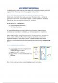
Normal vascular tone regulation by the endothelium
i) Endothelial NO synthase (eNOS) catalyzes oxidation of L-arginine → NO +
L-citrulline. NO diffuses across cell membranes and activates soluble guanylate cyclase
(sGC), which converts GTP to cGMP (which causes smooth muscle relaxation and
vasodilatation by lowering intracellular Ca²⁺, by efflux). There is also inducible NOS (iNOS),
induced by LPS, IFN-γ, IL-1 and other inflammatory signals in a wide range of cell types,
including macrophages and its primary role appears to be regulation of immune functions.
ii) Endothelins (ET-1, ET-2, ET-3) mediate endothelium-dependent
vasoconstriction and are potent mitogens for smooth muscle cells and fibroblasts and
trigger endothelial-mesenchymal transition. Thrombin, epinephrine, TNF, IFN-I, TGF-β, ANG-
II and hypoxia can all stimulate the release of ET-1 from endothelial cells.
iii) Procoagulant state
This state originates from the imbalance between coagulation and fibrinolysis: ↑ vWF,
increased platelet activation, ↑pro-atherogenic oxidized LDL and ↓ fibrinolysis. Platelets
are exposed to subendothelial connective tissue (after endothelial microinjury), which
promotes their aggregation.
2) Fibrosis
Fibrosis is prominent in the skin, lungs, GI tract, heart, tendon sheath, perifascicular tissue
surrounding skeletal muscle and in some endocrine organs such as the thyroid gland.
Fibrosis is type I collagen, fibronectin, proteoglycans and other structural macromolecules.
Endothelial-mesenchymal transition plays a critical role.
The lower esophagus is frequently involved, with prominent fibrosis of the lamina
propria and submucosa, characteristic vascular lesions and atrophy of the muscular layers.
Lower esophageal dysfunction leads to GERD. Chronic reflux is associated with esophageal
inflammation, ulcerations and stricture formation and may lead to Barrett’s esophagus.
In the lungs, patchy infiltration of the alveolar walls with CD8+, macrophages and
eosinophils is prominent in early disease. With disease progression, fibrosis and vascular
damage dominate the pathological picture in dSSc. Progressive thickening of the alveolar
septae results in obliteration of the air spaces and honeycombing as well as loss of
pulmonary blood vessels. This process impairs gas exchange and contributes to worsening
pulmonary hypertension. Extensive pulmonary fibrosis also may predispose to primary lung
carcinoma.
The electrical system of the heart may be involved, leading to conduction
disturbances. Also myocardium, pericardium are affected / fibrosed.
Epidemiology
Women = 5x men, 30-50yrs, declines after menopause, prevalence 100-300/million,
incidence: 18-20/million/year. African Americans have a higher incidence than Whites and
disease onset occurs at an earlier age, rare in children. African Americans are more likely to
have the diffuse cutaneous form of the disease with ILD and worse prognosis. The highest
prevalence is among Choctaw Native Americans (469/100.000). There’s an increased risk for
SSc patients to develop solid or hematologic malignancy.
Causes
Genetics: weak correlation with HLA-II (DRB1*11:04, DQA1*05:01, DQB1*03:01)
and non-HLA loci (PSORSIC1, Notch4). Associated genes outside HLA: PTPN22, which has
been associated with SLE, myasthenia gravis, vitiligo and Addison’s disease; Sox5, DNAse
1L3, PPAR-γ, CSK, CAV1, IL-1β, NLRP1, IRF5: the association of IRF5 with SSc is particularly
interesting in light of the potential pathogenic role of IFN-I in immune responses. The risk of
other autoimmune diseases, including SLE, RA, is also increased in 1st-degree relatives of SSc
patients. The results of SSc genetic studies to date can be broadly summarized as follows:
(1) the majority of SSc susceptibility loci are located in intronic gene regions that might
influence transcription of non-coding RNA or represent linkage disequilibrium with coding
or regulatory region genetic variants, (2) many SSc-associated genetic variants appear to be
involved in innate and adaptive immune responses, (3) they are largely shared with SLE and
other autoimmune and inflammatory diseases. The skin in SSc overexpresses genes
associated with fibroblasts, B-cells, endothelial cells.
Epigenetics: all three major types of epigenetic modifications (DNA
methylation, histone modifications including acetylation and methylation and expression of
noncoding [both long- and micro-] RNAs) have been described in SSc. MicroRNAs (miRNAs)
are a large family of 18-23 nucleotide noncoding RNAs that function as intracellular
regulators of gene expression. miRNAs can be detected in the circulation and they exert
biologic activities when incorporated into microvesicles.
Microchimerism: healthy women harbor circulating and occasionally tissue-
resident stem cells of fetal origin that persist for many years or even decades after
pregnancy. It has been suggested that the persistence of fetal cells in SSc patients might be
linked to disease pathogenesis through a GVHD–like response triggered by the fetal cells or
through a maternal (auto)immune response against the fetal cells. However, other evidence
suggests that microchimerism may have a protective effect in a variety of autoimmune and
other conditions.
Infections: CMV (anti-Scl70 recognize antigenic epitopes that are present on the
CMV-derived UL94 protein), EBV, ParvoB19.
Radiation therapy for malignant neoplasms: has been linked with the onset of de
novo SSc, as well as exacerbation of fibrosis in patients with preexisting SSc.
Pathophysiology
Hallmark: non-inflammatory, proliferative/obliterative vasculopathy of small arteries and
arterioles combined with interstitial fibrosis in target organs. Tissue inflammatory cells are
largely absent in long-standing SSc but may be prominent in early stages. Infiltrates are
located predominantly perivascularly and are composed of CD4+, B cells, DCs and
monocytes/macrophages. In the skin, the infiltrates are composed primarily of CD4+ and
monocytes, whereas CD8+ predominate in the lungs.
Three pathological processes:
,i) a small vessel non-inflammatory proliferative/obliterative vasculopathy
ii) autoimmunity/ inflammation
iii) fibrosis: the pathological accumulation of collagen (type I and other extracellular matrix)
in skin and other organs, probably comes after vasculopathy and autoimmunity.
1) Vasculopathy (endotheliopathy)
At the endothelial level, two pathways are predominantly activated, the inflammatory and
the microthrombotic one. Pathogenesis follows a chronological progression, with the
inflammatory pathway (endothelial activation and subsequent damage with apoptosis)
being predominant in the early phase and the microthrombotic pathway coming later. It’s
not a vasculitis as the vessel walls are not infiltrated (sometimes the adventitia might be,
though) and it’s not necessarily thrombotic (there’s not acute thrombus), but progressively
obliterative due to intimal proliferation.
Endothelial cell injury and apoptosis is thought to be the initial event in vascular
disease. Vascular injury causes activation of endothelial cells, with increased expression of
VCAM-1 and E-selectin and altered secretion of vasoactive mediators (ex. ↓NO), followed
by platelet aggregation, activation of the thrombotic and fibrinolytic cascades and
generation of thrombin, with impaired fibrinolysis and increased levels of circulating vWF.
Activated endothelial cells display durable epigenetic alterations and increased propensity
to transdifferentiate into mesenchymal cells via endothelial-mesenchymal transition (EMT)
driven by TGF-β and Notch. Circulating endothelial cells are mature cells shed from blood
vessels and are normally present only in very low numbers but are increased in a variety of
vascular diseases, where they appear to be markers of vascular damage. Circulating
endothelial cells are increased in SSc, consistent with the known vascular injury. In contrast,
circulating endothelial progenitor cells arise from the bone marrow and contribute to
vascular repair.
Activated platelets release circulating microparticles, PF4 (=CXCL4), TxA2, PDGF,
epidermal growth factor, HMGB1, serotonin and TGF-β. These mediators promote
coagulation, potentiate vasoconstriction and directly stimulate fibroblast activation and
myofibroblast transdifferentiation. Factors involved in this injury include autoantibodies,
infections, cytotoxic T-cells and ROS. Autoantibodies include anti-endothelial cell (AECA),
anti-angiotensin II receptor (ATRA) and anti-endothelin type-A receptor (ETRA) antibodies.
Signs of vasculopathy: Raynaud’s phenomenon (typically precedes other
manifestations of SSc), telangiectasias, nailfold infarcts, PAH, digital tip pitting and ischemic
ulcers, GAVE (watermelon stomach), SRC.
Scleroderma microvascular disease is characterized by:
i) microvasculopathy
ii) vasospasm
iii) procoagulant state with thrombosis and fibrin deposition
iv) defective angiogenesis
i) Microvasculopathy
The target of the disease are not the capillaries, but the arterioles (bigger). The
characteristic vascular lesion of SSc, bland intimal proliferation in the small and medium-
sized arteries, results in luminal narrowing/obliteration. Precedes fibrosis and can be
detected in clinically affected as well as unaffected skin.
Histologically: characteristic neointimal lesion (proliferation of endothelial and
smooth muscle cells and collagen deposition in intima layer, result from proliferation and
migration of myointimal cells and local accumulation of collagen and other ECM
components). The vascular basement membranes are thickened and reduplicated,
, adventitial fibrosis, perivascular mononuclear cell infiltration, pericyte activation, distortion
and loss of capillary loops. Different cells may be responsible for generating neointimal
lesions, including activated pericytes, adventitial cells, resident fibroblasts, vascular smooth
muscle and endothelial cells.
ii) Vasospasm
Prominent functional abnormalities of the vascular endothelium include impaired
production of and responsiveness to endothelium-derived vasodilators such as NO
(decreased expression of eNOS), thrombomodulin, calcitonin gene-related peptide and
prostacyclins and increased release of vasoconstrictors such as ET-1. Raynaud phenomenon
represents an exaggerated form of the normal thermoregulatory cold response that is
largely mediated by α2c-adrenoreceptors on vascular smooth muscle cells. Activated
endothelial cells release ET-1, which in addition to vasoconstriction also promotes leukocyte
adhesion, vascular smooth muscle cell proliferation and fibroblast activation.
Normal vascular tone regulation by the endothelium
i) Endothelial NO synthase (eNOS) catalyzes oxidation of L-arginine → NO +
L-citrulline. NO diffuses across cell membranes and activates soluble guanylate cyclase
(sGC), which converts GTP to cGMP (which causes smooth muscle relaxation and
vasodilatation by lowering intracellular Ca²⁺, by efflux). There is also inducible NOS (iNOS),
induced by LPS, IFN-γ, IL-1 and other inflammatory signals in a wide range of cell types,
including macrophages and its primary role appears to be regulation of immune functions.
ii) Endothelins (ET-1, ET-2, ET-3) mediate endothelium-dependent
vasoconstriction and are potent mitogens for smooth muscle cells and fibroblasts and
trigger endothelial-mesenchymal transition. Thrombin, epinephrine, TNF, IFN-I, TGF-β, ANG-
II and hypoxia can all stimulate the release of ET-1 from endothelial cells.
iii) Procoagulant state
This state originates from the imbalance between coagulation and fibrinolysis: ↑ vWF,
increased platelet activation, ↑pro-atherogenic oxidized LDL and ↓ fibrinolysis. Platelets
are exposed to subendothelial connective tissue (after endothelial microinjury), which
promotes their aggregation.
2) Fibrosis
Fibrosis is prominent in the skin, lungs, GI tract, heart, tendon sheath, perifascicular tissue
surrounding skeletal muscle and in some endocrine organs such as the thyroid gland.
Fibrosis is type I collagen, fibronectin, proteoglycans and other structural macromolecules.
Endothelial-mesenchymal transition plays a critical role.
The lower esophagus is frequently involved, with prominent fibrosis of the lamina
propria and submucosa, characteristic vascular lesions and atrophy of the muscular layers.
Lower esophageal dysfunction leads to GERD. Chronic reflux is associated with esophageal
inflammation, ulcerations and stricture formation and may lead to Barrett’s esophagus.
In the lungs, patchy infiltration of the alveolar walls with CD8+, macrophages and
eosinophils is prominent in early disease. With disease progression, fibrosis and vascular
damage dominate the pathological picture in dSSc. Progressive thickening of the alveolar
septae results in obliteration of the air spaces and honeycombing as well as loss of
pulmonary blood vessels. This process impairs gas exchange and contributes to worsening
pulmonary hypertension. Extensive pulmonary fibrosis also may predispose to primary lung
carcinoma.
The electrical system of the heart may be involved, leading to conduction
disturbances. Also myocardium, pericardium are affected / fibrosed.