Pathology: mechanisms of kidney injury
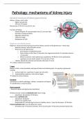
General structure of kidney parenchyma
Kidney = brown, red in color
- Highly vascularized
- 25% van CO output
Normal size: 10 X 6,5 X 3 cm (10-11 cm)
Function of kidney:
- Plasma filtration → concentrated urine (1L urine per day)
- Excretion of waste products
- Regulation blood flow
- Endocrine organ
- Metabolism of VitD
Nephrons and blood vessels
Nephron= structural and functional unit of the kidney, consists of the glomerulus + entire loop
- Bowman capsule: where filtration occurs
- Renal artery with glomerulus:
o Direct branch of abdominal aorta
o Enters kidney at the hilum → branches into segmental arteries → interlobar arteries
→ arterioles
o Afferent arteriole: enters glomerulus (capillaries) → efferent, forms peritubular
capillaries around all the tubules → renal veins → VCI
- Tubules: absorb the nutrients and secretes the toxins → urine
→ 1 million nephrons per kidney
2 types
Biopsy in clinic: cortex (medulla: only loop of Henle and collecting duct → majority is glomerular
related)
1. Cortical nephrons: superficial in upper part of cortex
2. Juxtamedullary nephrons: mid cortical region
→ related to certain diseases
Functions
Overvieuw on slide→ pathology = defects, mechanism of disease
→ ultrafiltration is mean function
Renal corpuscle
= bowman capsule and glomerulus (capillaries)
All blood vessels: lined by endothelial cells (inner lining)
- Specialized: fenestrated → filtration
Outer lining: epithelial cells
- Specialized: podocytes
- Holds on to capillary wall
- Responsible for maintaining structure of capillary lumen + have slit like spaces → filtration
Parietal epithelial cells: lines the Bowman’s space
- At tubular pole → become columnar, forms brush border → becomes proximal tubule cells
, Mesangium (connective tissue) connects the cells above
- Destroys foreign bodies of immune complexes
- Phagocytic action
Two pole ends: 1: vascular pole (where arterioles enters)
- Juxtaglomerular region: macula densa, juxtamedullary cells
o High profile filtration function
2: Tubular pole: proximal tubule
Chronic kidney disease
can be silent (prerenal – renal -postrenal) → late diagnosed
Global burden:
- Extremely common in the last 15 years
- Because of better diagnostic ways → more recognition of disease
- High mortality → important to diagnose early
RF:
- Elderly with comorbidities (diabetes, is a pandemic)
- Women
- Auto-immune diseases (lupus most common)
EU burden:
- 1/3 is at risk
- Mostly asymptomatic
1. Handling of kidney biopsy
Indications for renal biopsy
A. Unexplained acute of rapidly progressive renal failure
Certain parameters → see other lessons (decides grade of progression)
B. Nephrotic syndrome and non-nephrotic proteinuria
C. Persistent glomerular hematuria
D. Systemic diseases
E. Renal allograft dysfunction (tx)
Most common: auto-immune diseases
Handling
2 cores, 1-2 cm → we need mostly cortex, glomerular corpuscles (most
diseases: glomerular related)
Identify cortex or medulla under microscope
- Red points: not fixed tissue, blood gets stuck in glomeruli
Divide tissue: 1 glomerulus for fluorescence, one for EM, rest for
microscopy
Fixatives:
- Light microscopy: formaldehyde, maintains the architecture of the tissue
- Immunofluorescence: specialized microscopy, for identifying immunocomplex deficits → no
fixator: fresh frozen tissue
- EM: ultrastructural components of kidney, glutaraldehyde