Zelfstudie
The primer to the nervous system
Structure and function of the nervous system
- General function: reception and processing of sensory information (internal and external environment)
- 2 systems work together
Central NS: brain & spinal cord
. Brain protected by the skull
. Brain has direct connection with spinal cord (protected by vertebral column)
Peripheral NS: nerves (lie outside of CNS)
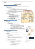
- Figure
Black: how CNS communicates with somatic NS and autonomic NS
Red: how CNS receives sensory info
- 3 specific functions
1. Receiving sensory input
. Via receptors in skin and other organs
. Generates nerve signals: from PNS -> CNS
2. Information processing and integration (CNS)
. Reviews, stores information as memories
. Creates appropriate motor response
3. CNS generates motor output
. Nerve signals: CNS -> PNS -> muscles/glands
Nervous tissue
- Has 2 types of cells
Neurons
. Function: transmission of nerve impulses between parts of NS
Neuroglia/glial cells
. Function: supporting and nourishing neurons
. Way more neuroglia than neurons in brain
. Myeline formed from membranes of tightly spiraled neuroglia
Shwann cells (PNS): gaps between = node of Ranvier
Oligodendrocytes (CNS)
. Microglia: phagocytic cells that help remove bacteria
. Astrocytes: metabolic and structural support directly to neurons
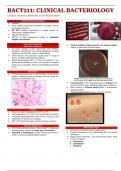
- Figure -> B) in CNS, some interneurons have a short axon that is not covered by myeline
Neuron anatomy
- 1. Sensory neurons
Function: signals from receptor to CNS
Sensory receptors: see change in environment
- 2. Interneurons
Function: sum up all the info received and communicates with motor neuron
Signals from sensory neurons or other interneurons
Lies within the CNS
- 3. Motor neurons
Function: takes nerve impulses away from the CNS to effector (muscle/organ/gland)
Effectors: carry out our responses to environmental changes
- Structure
Cell body: nucleus and organelles
Dendrites: receives signal from sensory neurons/receptors
Axon: conducts nerves signals/impulses received from the dendrites
. Nerve fiber: individual axon -> collectively: nerve
. Sensory neurons
Long axon carries signals from dendrites associated with sensory receptor to CNS
Axon interrupted by cell body
. Interneurons/motor neurons
1
, Zelfstudie
Multiple dendrites carry signals to cell body
Then axon conducts nerve signals away from the cell body
- Myeline around axons
Myeline: shwann or oligod. Wrap their membranes around axon
. Not all axons have myeline (shorts ones don’t have any)
Nodes of Ranvier: free spots without myeline
Function: transmissions peed, insulator, nerve regeneration PNS (new fiber growth when axon breaks)
. MS (multiple sclerosis): myeline breaks down -> difficulty with transmitting signals
Gray matter in CNS: no myeline
White matter in CNS: a lot of myeline
Neuron physiology
- In the past: only excised (taken from the body) neurons could be studied
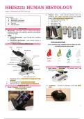
Resting Potential (see doc for foto)
- Like a battery: energy by separating positive and negative ions
Potential energy: used for work
- Resting potential: resting neuron has potential energy because plasma membrane is polarized
Polarizing: positive ions outside the cell, negative ions in the cell
- During action potential: Cell reaches threshold -> depolarisation -> repolarisation
- Positive outside (Na+)
Rest: membrane permeable to K+, not to Na+
K+ goes from inside the cell to the outside -> even more positive outside now
- Negative outside
Negative proteins and other molecules -> to big to diffuse
Action Potential
- Energy measured in volt (V) -> normally a cells has 0.070 V or 70 millivolts (mV)
Is a negative number (comparing negative inside to positive outside)
- Neurons must maintain their resting potential to work
By pushing Na+ out (more positive outside) of the cell and K+ into the cell (less positive outside)
- Action potential: conduction of nerve signals
In the axons of neurons
All or nothing event with threshold, only takes 3-4 ms
Ex: pushing a needle in your arm
. Pushing harder will not make the action potential stronger
. But! There will be more action potentials -> pain perceived as more intense
- Voltage-gated channels (protein channels)
Specific for Na ions
In plasma membrane of axon
- Action potential: Na+ gate opens -> Na+ into the cell -> inside more positive than outside (depolarisation: -70 mV
-55 mV -35 mV) -> Na+ gate closes -> K+ gate opens -> K+ to the outside of the cell -> inside becomes
negative again (repolarisation: ) -> sodium-potassium pomp: K+ to the inside & Na+ to the outside (completion)
Propagation of an Action Potential
- Unmyelinated fibers: slow action potential (1 m/s, small fibers)
Each axon must be stimulated
Action potential stimulates adjacent part of the axon membrane
- Myelinated fibers: fast action potential (100 m/s, thick fibers) = salutatory condition
Jumps from one node of Ranvier to the other
- Self-propagating: each AP generates another AP along the entire length of the axon
Conduction is all or nothing event
Intensity: how many AP in a given time
- Refractory period after conducting and AP -> ensures one-way direction
2
The primer to the nervous system
Structure and function of the nervous system
- General function: reception and processing of sensory information (internal and external environment)
- 2 systems work together
Central NS: brain & spinal cord
. Brain protected by the skull
. Brain has direct connection with spinal cord (protected by vertebral column)
Peripheral NS: nerves (lie outside of CNS)
- Figure
Black: how CNS communicates with somatic NS and autonomic NS
Red: how CNS receives sensory info
- 3 specific functions
1. Receiving sensory input
. Via receptors in skin and other organs
. Generates nerve signals: from PNS -> CNS
2. Information processing and integration (CNS)
. Reviews, stores information as memories
. Creates appropriate motor response
3. CNS generates motor output
. Nerve signals: CNS -> PNS -> muscles/glands
Nervous tissue
- Has 2 types of cells
Neurons
. Function: transmission of nerve impulses between parts of NS
Neuroglia/glial cells
. Function: supporting and nourishing neurons
. Way more neuroglia than neurons in brain
. Myeline formed from membranes of tightly spiraled neuroglia
Shwann cells (PNS): gaps between = node of Ranvier
Oligodendrocytes (CNS)
. Microglia: phagocytic cells that help remove bacteria
. Astrocytes: metabolic and structural support directly to neurons
- Figure -> B) in CNS, some interneurons have a short axon that is not covered by myeline
Neuron anatomy
- 1. Sensory neurons
Function: signals from receptor to CNS
Sensory receptors: see change in environment
- 2. Interneurons
Function: sum up all the info received and communicates with motor neuron
Signals from sensory neurons or other interneurons
Lies within the CNS
- 3. Motor neurons
Function: takes nerve impulses away from the CNS to effector (muscle/organ/gland)
Effectors: carry out our responses to environmental changes
- Structure
Cell body: nucleus and organelles
Dendrites: receives signal from sensory neurons/receptors
Axon: conducts nerves signals/impulses received from the dendrites
. Nerve fiber: individual axon -> collectively: nerve
. Sensory neurons
Long axon carries signals from dendrites associated with sensory receptor to CNS
Axon interrupted by cell body
. Interneurons/motor neurons
1
, Zelfstudie
Multiple dendrites carry signals to cell body
Then axon conducts nerve signals away from the cell body
- Myeline around axons
Myeline: shwann or oligod. Wrap their membranes around axon
. Not all axons have myeline (shorts ones don’t have any)
Nodes of Ranvier: free spots without myeline
Function: transmissions peed, insulator, nerve regeneration PNS (new fiber growth when axon breaks)
. MS (multiple sclerosis): myeline breaks down -> difficulty with transmitting signals
Gray matter in CNS: no myeline
White matter in CNS: a lot of myeline
Neuron physiology
- In the past: only excised (taken from the body) neurons could be studied
Resting Potential (see doc for foto)
- Like a battery: energy by separating positive and negative ions
Potential energy: used for work
- Resting potential: resting neuron has potential energy because plasma membrane is polarized
Polarizing: positive ions outside the cell, negative ions in the cell
- During action potential: Cell reaches threshold -> depolarisation -> repolarisation
- Positive outside (Na+)
Rest: membrane permeable to K+, not to Na+
K+ goes from inside the cell to the outside -> even more positive outside now
- Negative outside
Negative proteins and other molecules -> to big to diffuse
Action Potential
- Energy measured in volt (V) -> normally a cells has 0.070 V or 70 millivolts (mV)
Is a negative number (comparing negative inside to positive outside)
- Neurons must maintain their resting potential to work
By pushing Na+ out (more positive outside) of the cell and K+ into the cell (less positive outside)
- Action potential: conduction of nerve signals
In the axons of neurons
All or nothing event with threshold, only takes 3-4 ms
Ex: pushing a needle in your arm
. Pushing harder will not make the action potential stronger
. But! There will be more action potentials -> pain perceived as more intense
- Voltage-gated channels (protein channels)
Specific for Na ions
In plasma membrane of axon
- Action potential: Na+ gate opens -> Na+ into the cell -> inside more positive than outside (depolarisation: -70 mV
-55 mV -35 mV) -> Na+ gate closes -> K+ gate opens -> K+ to the outside of the cell -> inside becomes
negative again (repolarisation: ) -> sodium-potassium pomp: K+ to the inside & Na+ to the outside (completion)
Propagation of an Action Potential
- Unmyelinated fibers: slow action potential (1 m/s, small fibers)
Each axon must be stimulated
Action potential stimulates adjacent part of the axon membrane
- Myelinated fibers: fast action potential (100 m/s, thick fibers) = salutatory condition
Jumps from one node of Ranvier to the other
- Self-propagating: each AP generates another AP along the entire length of the axon
Conduction is all or nothing event
Intensity: how many AP in a given time
- Refractory period after conducting and AP -> ensures one-way direction
2