HHIS221: HUMAN HISTOLOGY
Lesson 1 | Microscope and Cell & Cell Cycle
2) Objective Lens - metal cylinders attached below the
nosepiece and contains especially ground and polished
Topic Outline:
● Microscope lenses. – Most sensitive and expensive part
● Microscopic Techniques ● Scanning Objective (Red) - 4x
● Care of the Microscope ● LPO (Yellow) - 10x
● The Cell ● HPO (Blue) - 40x
● The Cell Cycle ● OIO (White) - 100x
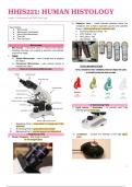
Microscope
● Microscope - instrument used for viewing small objects,
including microbes, can magnify a specimen and optically
resolve fine detail.
● Types:
1) Simple Microscope - uses a single lens to magnify
the object.
2) Compound Microscope - uses several lenses to
magnify the object. TOTAL MAGNIFICATION
Parts of a Compound Microscope
II. Illuminating Parts
1) Light source / Illuminator - to provide even, high
intensity light (provide light)
I. Magnifying Parts – responsible for enlargement of the
specimen
II. Illuminating Parts – gives light to the sample
III. Mechanical Part – provide support and adjustment
I. Magnifying Parts 2) Condenser - controls the intensity of the light (glass
1) Ocular / Eyepiece - final image is viewed lens)
- 10-15x magnification
1
, 3) Iris Diaphragm - controls the light reaching the object. Care of the Microscope
(controls the amount of light passing through the
condenser)
The Cell
1) Nucleus
● Structure:
III. Mechanical Parts
– Large structure enclosed within a double
1) Draw Tube - situated on the top of the body tube ,holds
membrane; contains chromatin, nucleolus, and
eyepiece
nucleoplasm
2) Body Tube - separates the objective and the eyepiece
● Major Function:
3) Arm - a curved or slanted part that is held while carrying
– Houses the DNA that serves as the genetic material
the microscope.
for directing protein synthesis.
4) Base - bottommost portion that supports the entire/lower
2) Nucleolus
microscope
● Structure:
5) Revolving Nosepiece /Turret- where the objectives are
– Large, prominent structure within the nucleus
attached
● Major Function:
6) Mechanical Stage - platform where object to be
– for synthesis of ribosomes
examined is placed (clip, slide holder, and aperture)
3) Nuclear Envelope
7) Coarse Adjustment Knob - used for initial focusing
● Structure:
8) Fine Adjustment Knob - for fine tuning/sharp focusing
– Double membrane boundary between cytoplasm
and nuclear contents; continuous with rough
NOTE: You will use the coarse adjustment knob only on
endoplasmic reticulum
Scanning objective and LPO only, once you are using HPO
● Major Function:
and OIO you can only use fine adjustment knob.
– Separates nucleus from cytoplasm
4) Cytoplasm
Microscopic Techniques
● Responsible for many cellular processes
I. Bright-field microscopy - allows light rays to pass
5) Cytosol
directly through to the eye without being deflected by an
● Provides support for organelles; serves as the viscous
intervening opaque plate in the condenser
fluid medium through which diffusion occurs
II. Dark-field microscopy - to observe transparent
6) Organelles
microorganisms
● Carry out specific metabolic activities of the cell
III. Phase-contrast microscopy - a microscope that
7) Endoplasmic Reticulum
differentiates transparent protoplasmic structures
● consists of a network of membranous tubules and
without staining and killing them (identification of viable
sacs called cisternae
microorganisms)
8) Rough Endoplasmic Reticulum
IV. Fluorescence microscopy - a microscope that
● Have attached ribosomes
involves the use of differential dyes/fluorochromes and
● synthesis of secretory proteins, cell membrane protein
immunofluorescence techniques.
and organelle protein
V. Polarizing microscopy - equipped with specialized
9) Smooth Endoplasmic Reticulum
filters which orient light in a manner which can be used
● Without attachment of ribosomes
to identify and enhance characteristics such as
● synthesis of lipid
birefringence exhibited by certain substances.
● glycogen metabolism in the liver cells
VI. Electron microscopy - attains extremely high
● store calcium
resolution using an electron beam instead of a beam of
light to illuminate the object of study.
a) Transmission Electron Microscopy (TEM)
b) Scanning Electron Microscopy (SEM)
2
Lesson 1 | Microscope and Cell & Cell Cycle
2) Objective Lens - metal cylinders attached below the
nosepiece and contains especially ground and polished
Topic Outline:
● Microscope lenses. – Most sensitive and expensive part
● Microscopic Techniques ● Scanning Objective (Red) - 4x
● Care of the Microscope ● LPO (Yellow) - 10x
● The Cell ● HPO (Blue) - 40x
● The Cell Cycle ● OIO (White) - 100x
Microscope
● Microscope - instrument used for viewing small objects,
including microbes, can magnify a specimen and optically
resolve fine detail.
● Types:
1) Simple Microscope - uses a single lens to magnify
the object.
2) Compound Microscope - uses several lenses to
magnify the object. TOTAL MAGNIFICATION
Parts of a Compound Microscope
II. Illuminating Parts
1) Light source / Illuminator - to provide even, high
intensity light (provide light)
I. Magnifying Parts – responsible for enlargement of the
specimen
II. Illuminating Parts – gives light to the sample
III. Mechanical Part – provide support and adjustment
I. Magnifying Parts 2) Condenser - controls the intensity of the light (glass
1) Ocular / Eyepiece - final image is viewed lens)
- 10-15x magnification
1
, 3) Iris Diaphragm - controls the light reaching the object. Care of the Microscope
(controls the amount of light passing through the
condenser)
The Cell
1) Nucleus
● Structure:
III. Mechanical Parts
– Large structure enclosed within a double
1) Draw Tube - situated on the top of the body tube ,holds
membrane; contains chromatin, nucleolus, and
eyepiece
nucleoplasm
2) Body Tube - separates the objective and the eyepiece
● Major Function:
3) Arm - a curved or slanted part that is held while carrying
– Houses the DNA that serves as the genetic material
the microscope.
for directing protein synthesis.
4) Base - bottommost portion that supports the entire/lower
2) Nucleolus
microscope
● Structure:
5) Revolving Nosepiece /Turret- where the objectives are
– Large, prominent structure within the nucleus
attached
● Major Function:
6) Mechanical Stage - platform where object to be
– for synthesis of ribosomes
examined is placed (clip, slide holder, and aperture)
3) Nuclear Envelope
7) Coarse Adjustment Knob - used for initial focusing
● Structure:
8) Fine Adjustment Knob - for fine tuning/sharp focusing
– Double membrane boundary between cytoplasm
and nuclear contents; continuous with rough
NOTE: You will use the coarse adjustment knob only on
endoplasmic reticulum
Scanning objective and LPO only, once you are using HPO
● Major Function:
and OIO you can only use fine adjustment knob.
– Separates nucleus from cytoplasm
4) Cytoplasm
Microscopic Techniques
● Responsible for many cellular processes
I. Bright-field microscopy - allows light rays to pass
5) Cytosol
directly through to the eye without being deflected by an
● Provides support for organelles; serves as the viscous
intervening opaque plate in the condenser
fluid medium through which diffusion occurs
II. Dark-field microscopy - to observe transparent
6) Organelles
microorganisms
● Carry out specific metabolic activities of the cell
III. Phase-contrast microscopy - a microscope that
7) Endoplasmic Reticulum
differentiates transparent protoplasmic structures
● consists of a network of membranous tubules and
without staining and killing them (identification of viable
sacs called cisternae
microorganisms)
8) Rough Endoplasmic Reticulum
IV. Fluorescence microscopy - a microscope that
● Have attached ribosomes
involves the use of differential dyes/fluorochromes and
● synthesis of secretory proteins, cell membrane protein
immunofluorescence techniques.
and organelle protein
V. Polarizing microscopy - equipped with specialized
9) Smooth Endoplasmic Reticulum
filters which orient light in a manner which can be used
● Without attachment of ribosomes
to identify and enhance characteristics such as
● synthesis of lipid
birefringence exhibited by certain substances.
● glycogen metabolism in the liver cells
VI. Electron microscopy - attains extremely high
● store calcium
resolution using an electron beam instead of a beam of
light to illuminate the object of study.
a) Transmission Electron Microscopy (TEM)
b) Scanning Electron Microscopy (SEM)
2