Cell death in thophysiology - Summary
1. Introduction [2]
❥ Stress is a broad term and can both mean acute and chronic stress
❣ Acute stress is short-lived and usually not that bad long-term
❣ Chronic stress is long-term and can have lasting effects on the body
❥ Stress can refer to an experience influenced by emotions and thoughts but can also refer to cellular
stress which is a measurable phenomenon
❥ Cellular stress can be influenced by environmental factors: DNA damage, pathogens, heat, metabolic
stress, oxidative stress and senescence
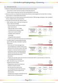
❥ How does the body deal with cellular stress?
❣ When cellular stress is still low the body first
tries to repair the damage
⤷ This can be done via numerous cellular
repair mechanisms
⧙ DNA damage response (DDR)
⧙ Unfolded protein response (UPR)
⧙ Heat shock response (HSR)
⧙ Oxidative stress response
⧙ Autophagy
⧙ Integrated stress response
⤷ If this is successful the body returns to homeostasis
❣ If cellular repair is unsuccessful the body
goes to the next step: cell death of cellular
senescence
⤷ If the cell can’t fix the damage it has
accumulated due to cellular stress it can
either go into a dormant mode or kill itself
⤷ If it goes in a dormant mode we speak of
senescence: cells in this state keep the
cellular structure but are metabolically
inactive (they still do have some
functions!)
⤷ If it goes into suicide mode, the cell will try to dismantle itself in a way that does not harm the
surrounding cells and structure
❣ After cell death occurs, the body will
phagocyte all the remains
⤷ Phagocytosis means the cell will eat
whatever remnants remain
⤷ Not only does this clean up the
surrounding area but it also leaves the
opportunity to recycle organels and
biomolecules
⤷ If phagocytosis happens limitedly then the
body can return to homeostasis
Page 1 |
, Cell death in thophysiology - Summary
❣ If phagocytosis occurs massively the body
goes into inflammation
⤷ The body can either resolve this
inflammation on its own and return to
homeostasis
⤷ Or the body can fail at resolving the
inflammation and disease can occur such
as cancer, excessive necrosis,
autoimmunity, infection, septic shock,
organ failure, etc.
❥ Another way to visualise how the body progresses through cellular stress is the triangle of death:
❥ The body constantly renews itself
❣ The body consists of 100 trillion cells
❣ 1-5 million cells die every second
❣ So clearly the body needs to constantly make new cells otherwise a human wouldn’t live very long
❣ The human genome consists of 6 billion nucleotides that get copied 5 million times per second
❥ So there is a clear delicate balance between cellular death and cellular renewal
❥ Disrupting this balance in either side can give issues
❣ Too much cell death and you get diseases with phenotypes such as organ injury and degeneration
❣ Too much cell renewal and you get cancer
❥ Unfortunately this balance does fall into disbalance often leading to one or the other scenarios
Page 2 |
, Cell death in thophysiology - Summary
2. Molecular mechanisms of cellular stress and cell death [15]
Exam questions:
❥ MCQ
❥ Terminology
2.1. DNA damage response (DDR)
2.1.A. Types of DNA damage
① Overview
1. Replication errors
❣ DNA polymerase has an error rate of 1 in 106
⤷ This equates to 3000 errors per cell division
⤷ When we take the amount of cells that are made every second into consideration (5∙106 new cells
per second) then we get 15 billion repairs per second in the human body
❣ Replication errors lead to base mismatches and bulges in the DNA strand
❣ Replication errors are repaired by: mismatch repair (MMR)
2. Oxidative damage
❣ Caused by reactive oxygen species: highly reactive oxygen-containing molecules that can react with
different biomolecules such as nucleotides, lipids and amino acids leading to the formation of other
radicals that can further disrupt cellular structures such as cellular membranes, DNA and proteins
❣ ROS cause single-strand break (SSB)
❣ Oxidative damage can be fixed by: base excision repair
3. Alkylation and hydrolysis
❣ Alkylation and hydrolysis cause single base damage
❣ Alkylation and hydrolysis can be repaired by: base excision repair
4. Ionizing radiation
❣ Ionizing radiation include gamma rays and X-rays
❣ These can cause double-strand breaks (DBS)
❣ Damage by ionizing radiation can be repaired by: non-homologous end joining (NHEJ) and
homology-directed repair (HDR)
5. Chemotherapeutics
❣ Can lead to interstrand crosslinks
❣ Can be repaired by: NHEJ, HDR and nucleotide excision repair (NER)
6. UV light and radicals
❣ Can lead to intrastrand crosslinks (e.g. thymine dimers) and bulky adducts
❣ Can be repaired by: NER
Page 3 |
, Cell death in thophysiology - Summary
② Oxidative damage to DNA
❥ Electron biology can be very impactful in deciding if a cell lives or not
❥ Oxygen is important in electron biology due to its two unpaired electrons in split orbitals
❥ Because of this, oxygen is toxic and can be further oxidized into highly reactive oxygen species:
❥ These ROS can damage cellular structures by reacting with biomolecules and turning them into
radicals
❣ ROS can react with nucleotides to form oxidized nucleotides that result in mutations: guanine → 8-
oxoguanine → pairing with adenine (instead of cytosine) → 8-oxoG is excised to T → G→T transversion
❣ ROS can react with proteins leading to loss of function, unfolding and aggregation
❣ ROS can react with lipids leading to lipid peroxidation
③ Radiation damage to DNA
Page 4 |