CEREBROVASCULAR
ANATOMY
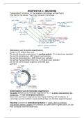
During early embryonic development, six paired aortic arches form
sequentially between the ventral aorta (emerging from the heart) and the
dorsal aorta. The first aortic arch contributes to the maxillary artery. The
second aortic arch gives rise to parts of the hyoid and stapedial arteries.
The third aortic arch is crucial for carotid artery development. It forms
part of the future common carotid artery and the proximal portion of the
internal carotid artery. The internal and external carotid arteries originate
from this system through regression.
THE CAROTID ARTERY SYSTEM
Common carotid arteries:
o The third aortic arch contributes to the proximal
portions of the common carotid arteries bilaterally.
o The ventral aorta between the heart and the third arch becomes the proximal portion of the
common carotid artery.
Internal carotid arteries:
o The dorsal aorta segment distal to the third arch forms the internal carotid arteries.
o These arteries grow cranially to supply the developing brain.
External carotid arteries:
o The external carotid arteries arise from sprouting branches of the ventral aorta near the third
aortic arch.
Regression of the first and second aortic arches ensures proper routing of blood flow, with their remnants
contributing to smaller arterial branches rather than the major carotid system. The third aortic arch and
portions of the dorsal aorta play a critical role in forming segments of the internal carotid artery (ICA). The ICA
can be divided into segments based on its path:
1. Cervical ICA
o The third aortic arch forms the proximal segment of the internal carotid artery, known as the
cervical segment.
o This segment extends from its origin at the common carotid artery bifurcation to the base of
the skull, where it enters the carotid canal.
2. Ascending intrapetrosal segment
o As the ICA enters the skull through the carotid canal in the temporal bone, it ascends
vertically within the petrous part of the temporal bone.
3. Horizontal intrapetrosal segment
o After ascending in the petrous bone, the ICA turns medially and takes a horizontal course
within the temporal bone, forming the horizontal intrapetrosal segment.
POSTERIOR DIVISION OF ICA
In the embryonic stage, the posterior division of the ICA contributes significantly to the development of the
posterior cerebral circulation, which is primarily supplied by the vertebrobasilar system in adults. In the fetal
stage, the posterior circulation initially derives from the ICAs through the posterior division. The key features
include:
1. Midline fusion of both posterior divisions of the ICA
The posterior divisions of the ICAs on both sides converge and fuse at the midline to form the
distal basilar artery.
Page | 1
, o This basilar artery develops as a single midline vessel, supplying the hindbrain (including
the cerebellum, brainstem, and posterior cerebral hemispheres).
o The vertebral arteries have not yet fully developed or taken over this role in the early
fetal stage.
2. Flow direction opposite to the adult situation
In the fetus, blood flows cranio-caudally, from the ICAs through the posterior communicating
arteries (PCOMs) into the fused basilar artery.
o This contrasts with the adult situation, where the flow is primarily caudo-cranial:
Blood ascends through the vertebral arteries and into the basilar artery to reach
the posterior cerebral circulation.
The cranio-caudal flow pattern in the fetal stage reflects the dependency of the posterior
circulation on the ICAs before the vertebral arteries assume their mature role.
The vertebral artery develops through a process involving the formation of
metameric vascular segments and their subsequent longitudinal fusion. In the
embryonic posterior cervical region, there are paired intersegmental arteries
that arise from the dorsal aorta. These arteries are segmental (metameric) and
supply blood to each somite (the embryonic precursors of vertebrae and
associated structures). During development, the cervical intersegmental
arteries begin to anastomose with one another longitudinally. The longitudinal
anastomosis forms a continuous vertebral artery on each side of the neck,
connecting multiple intersegmental arteries. Not all intersegmental arteries
persist. Segments that do not contribute to the longitudinal anastomosis
regress. This selective regression ensures a single, continuous vertebral artery
while eliminating redundant pathways.
The basilar artery, a major component of the posterior cerebral circulation,
forms through a process of midline fusion involving the vertebral arteries.
Transverse fusion refers to the process where the paired ventral longitudinal
neural arteries, running along the sides of the embryonic neural tube, connect
transversely through anastomoses. These transverse connections help to
establish early blood supply to the midline structures of the developing
hindbrain. As the vertebral arteries ascend cranially along the sides of the
cervical spinal cord and brainstem, they approach the midline. At the level of the medulla oblongata, the two
vertebral arteries join to form a single, midline basilar artery. The newly formed basilar artery runs along the
midline of the pons and gives rise to key branches, which are the anterior inferior cerebellar arteries (AICA),
pontine arteries, superior cerebellar arteries (SCA), and the posterior cerebral arteries (PCA).
In the adult circulation, the vertebrobasilar system becomes the dominant supply to the posterior circulation
instead of the PCOMs. The PCOMs regress in size and function, becoming thin collateral pathways in most
individuals.
ANTERIOR DIVISION OF ICA
The anterior division of the ICA gives rise to several critical arteries supplying the anterior and middle portions
of the brain. These vessels form part of the anterior cerebral circulation and contribute to perfusion of the
cortex, deep brain structures, and basal ganglia.
Middle cerebral artery (MCA): A terminal branch of the ICA, arising from the anterior division.
Anterior cerebral artery (ACA): The second terminal branch of the ICA.
Lenticulostriate arteries: Branches of the proximal MCA.
o Anterior choroidal artery: A small branch of the ICA, just proximal to its bifurcation.
o Perforators from the ACA: These small arteries arise from the ACA and include the recurrent
artery of Heubner
Recurrent artery of Heubner: Supplies the caudate nucleus, anterior internal capsule,
and putamen. It is an important artery for basal ganglia perfusion.
o Medial and lateral perforators from the MCA: Medial perforators are branches from the
MCA's proximal segment supply the medial portions of the basal ganglia and internal capsule.
Page | 2
, Lateral perforators are branches that supply the lateral portions of the basal ganglia and
parts of the white matter.
The ACA is divided into segments based on its course and branching pattern. The A1 segment is the initial part
of the ACA that extends from its origin at the ICA to the anterior communicating artery (ACOM). The A2
segment starts at the anterior communicating artery and extends along the medial surface of the brain,
looping around the corpus callosum. The ACOM connects the two ACAs at the midline, forming part of the
Circle of Willis.
Variations in the anatomy of the ACA and ACOM are common and may influence cerebral blood flow, the
development of collateral circulation, and susceptibility to certain vascular conditions.
Fenestrations of the ACOM: A fenestration refers to a segment of the artery that splits into two
parallel channels, creating a "window-like" structure within the vessel.
o Duplicated ACOM: When the ACOM forms two separate channels, each connecting the two
ACAs.
o Duplicated ACA (A1-A2): Refers to the presence of two separate A1 segments (proximal ACA)
or A2 segments (distal ACA), or both.
o Double duplication: Refers to the presence of two duplicated ACOMs or two duplicated ACA
segments (either A1 or A2).
o Triple duplication: Refers to the presence of three distinct vascular channels in either the
ACOM or ACA segments (A1 or A2).
Asymmetrical configuration of the ACA-A1 segment:
o Aplasia of A1: Complete absence of the A1 segment on one side. Collateral flow through the
ACOM is critical in this scenario.
o Hypoplasia of A1: A1 is underdeveloped or significantly smaller on one side. Blood flow is
predominantly provided by the contralateral ACA via the ACOM.
Fusion and ‘early bifurcation’ variations:
o Azygos configuration: The two A2 segments fail to separate and fuse into a single midline
artery, referred to as an azygos anterior cerebral artery. This single artery supplies both
medial hemispheres.
o Pericallosal and callosomarginal artery origin from ACOM: Normally, the pericallosal and
callosomarginal arteries arise as branches of the A2 segment of the ACA. In some cases, one
or both of these arteries can arise directly from the ACOM, bypassing the A2 segment.
The middle cerebral artery (MCA) is the continuation of the ICA after it passes through the Circle of Willis.
After leaving the Circle of Willis, the MCA bifurcates into two major branches:
1. Superior division: Supplies the frontal lobe, lateral portions of the motor and sensory cortices.
2. Inferior division: Supplies the lateral temporal lobe and the parietal lobe's posterior regions, including
areas involved in speech and language.
M1 refers to the proximal portion of the MCA, also known as the horizontal segment. It begins at the ICA
bifurcation (where the ICA branches into the MCA and ACA). M2 is the segment of the MCA that arises after
the M1 segment passes into the Sylvian fissure and bifurcates into the superior division and inferior division.
Despite its relatively predictable course and branching pattern, the MCA can exhibit several variations both in
its anatomy and in its relationship with other cerebral vessels.
Aneurysms are often located in the ACOM, especially when there is asymmetry. If the neck of the aneurysm is
small, coiling is favourable. If the neck is very wide, the coil can move down and occlude the artery. This can be
prevented with balloon assisted coiling, which prevents the coils from going into the vessel. A second option is
stent assisted coiling. This is an open stent, through which a small microcatheter fits to put coils in the
aneurysm. Having an hemorrhage gives a major drawback. Now, it is not safe to give patient anticoagulation
treatment. Instead of putting in stent, they try to coil in the acute stage and leave a little bit open. In the long
term, it will regrow and the coils will be compressed. After a few months, additional treatment is needed.
There is a very short and wide aneurysm left, which is hard to resolve. Now, they have to place the stent from
left to right (not from bottom to either left or right), which can be done by using the PCOM.
PET IMAGE PROCESSING &
QUANTIFICATION
Page | 3
, The principles of PET imaging are:
Annihilation process: Positrons (𝛽+) emitted from the radioactive isotope collide with electrons (𝑒−) in
the tissue, leading to their annihilation. This annihilation event produces two photons, each with an
energy of 511 keV, emitted in opposite directions.
Detection mechanism: These 511 keV photons are
detected by the PET scanner. Detection relies on a
system of opposing detectors that capture the
coincident photons. Coincidence detection ensures only
photons from true annihilation events are considered,
aiding in image accuracy.
Positron behaviour in tissue: Positrons travel a short
distance (~1.1 mm on average in tissue) before
annihilation, losing energy through scattering
interactions.
Isotope stability: The isotopes used are positron-
emitting and unstable, eventually decaying as part of the process that enables imaging.
PET is highlighted as an essentially quantitative imaging technique that can measure changes in metabolism
and function before any structural changes occur in the tissues. Applications of molecular imaging with PET:
Diagnosis and staging: PET imaging helps identify and stage diseases, such as cancer, by detecting
metabolic activity and functional changes in tissues.
Biological characterization: Provides detailed insights into biological processes, helping understand
the metabolic pathways and functional states of tissues.
Therapy response evaluation: PET can monitor how a treatment is working by observing changes in
tissue metabolism or function after the initiation of therapy.
Restaging: Useful for reevaluating the disease's progression or recurrence after a period of treatment
or remission.
Personalized medicine: Different radiopharmaceutical tracers target specific metabolic pathways,
allowing customization of imaging and treatment strategies to individual patient needs.
METABOLIC PET IMAGING
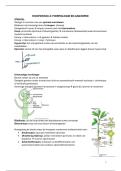
FDG is an analogue of glucose where 1 oxygen is replaced
by 18F. It behaves almost the same as normal glucose.
Tumours use glucose less efficient than healthy tissue.
They often switch to anaerobe glucose dissimilation,
which gives less ATP than aerobe dissimilation. FDG is
transported into tissue by the same transporters as
glucose, but with different Michaelis-Menten parameters.
The first step of glycolysis is using hexokinase to create glucose-6-P or FDG-6-P. Then, FDG stops, because FDG-
6-P can’t be broken down by the body. However, it does decay. Also, it can be converted back (k4), but this
process takes hours, and is thus not seen on the PET image.
The standardized uptake value (SUV) is a key metric for PET image quantification. The formula is:
Activity concentration at time t
SUV = ' .
Injected dose / Patient s body weight
Numerator: Represents the activity concentration (Bq/ml) measured in a region of interest within the
PET image.
Denominator: Normalizes this value by the injected dose (MBq) adjusted for patient body weight (kg).
It provides a standardized, semi-quantitative metric that allows comparison of tracer uptake across different
patients, scans performed at different times, and various clinical scenarios or research settings.
Static PET imaging captures data within a predetermined time interval after tracer injection, often when tracer
distribution has reached a relatively stable state. A common interval is 60–90 minutes post-injection. It
represents the average activity concentration during the imaging session. While static PET is simpler than
dynamic imaging, it still requires knowledge of optimal uptake time, ensuring the tracer is evenly distributed in
the area of interest. Static imaging does not provide dynamic information, meaning it cannot decompose the
Page | 4
ANATOMY
During early embryonic development, six paired aortic arches form
sequentially between the ventral aorta (emerging from the heart) and the
dorsal aorta. The first aortic arch contributes to the maxillary artery. The
second aortic arch gives rise to parts of the hyoid and stapedial arteries.
The third aortic arch is crucial for carotid artery development. It forms
part of the future common carotid artery and the proximal portion of the
internal carotid artery. The internal and external carotid arteries originate
from this system through regression.
THE CAROTID ARTERY SYSTEM
Common carotid arteries:
o The third aortic arch contributes to the proximal
portions of the common carotid arteries bilaterally.
o The ventral aorta between the heart and the third arch becomes the proximal portion of the
common carotid artery.
Internal carotid arteries:
o The dorsal aorta segment distal to the third arch forms the internal carotid arteries.
o These arteries grow cranially to supply the developing brain.
External carotid arteries:
o The external carotid arteries arise from sprouting branches of the ventral aorta near the third
aortic arch.
Regression of the first and second aortic arches ensures proper routing of blood flow, with their remnants
contributing to smaller arterial branches rather than the major carotid system. The third aortic arch and
portions of the dorsal aorta play a critical role in forming segments of the internal carotid artery (ICA). The ICA
can be divided into segments based on its path:
1. Cervical ICA
o The third aortic arch forms the proximal segment of the internal carotid artery, known as the
cervical segment.
o This segment extends from its origin at the common carotid artery bifurcation to the base of
the skull, where it enters the carotid canal.
2. Ascending intrapetrosal segment
o As the ICA enters the skull through the carotid canal in the temporal bone, it ascends
vertically within the petrous part of the temporal bone.
3. Horizontal intrapetrosal segment
o After ascending in the petrous bone, the ICA turns medially and takes a horizontal course
within the temporal bone, forming the horizontal intrapetrosal segment.
POSTERIOR DIVISION OF ICA
In the embryonic stage, the posterior division of the ICA contributes significantly to the development of the
posterior cerebral circulation, which is primarily supplied by the vertebrobasilar system in adults. In the fetal
stage, the posterior circulation initially derives from the ICAs through the posterior division. The key features
include:
1. Midline fusion of both posterior divisions of the ICA
The posterior divisions of the ICAs on both sides converge and fuse at the midline to form the
distal basilar artery.
Page | 1
, o This basilar artery develops as a single midline vessel, supplying the hindbrain (including
the cerebellum, brainstem, and posterior cerebral hemispheres).
o The vertebral arteries have not yet fully developed or taken over this role in the early
fetal stage.
2. Flow direction opposite to the adult situation
In the fetus, blood flows cranio-caudally, from the ICAs through the posterior communicating
arteries (PCOMs) into the fused basilar artery.
o This contrasts with the adult situation, where the flow is primarily caudo-cranial:
Blood ascends through the vertebral arteries and into the basilar artery to reach
the posterior cerebral circulation.
The cranio-caudal flow pattern in the fetal stage reflects the dependency of the posterior
circulation on the ICAs before the vertebral arteries assume their mature role.
The vertebral artery develops through a process involving the formation of
metameric vascular segments and their subsequent longitudinal fusion. In the
embryonic posterior cervical region, there are paired intersegmental arteries
that arise from the dorsal aorta. These arteries are segmental (metameric) and
supply blood to each somite (the embryonic precursors of vertebrae and
associated structures). During development, the cervical intersegmental
arteries begin to anastomose with one another longitudinally. The longitudinal
anastomosis forms a continuous vertebral artery on each side of the neck,
connecting multiple intersegmental arteries. Not all intersegmental arteries
persist. Segments that do not contribute to the longitudinal anastomosis
regress. This selective regression ensures a single, continuous vertebral artery
while eliminating redundant pathways.
The basilar artery, a major component of the posterior cerebral circulation,
forms through a process of midline fusion involving the vertebral arteries.
Transverse fusion refers to the process where the paired ventral longitudinal
neural arteries, running along the sides of the embryonic neural tube, connect
transversely through anastomoses. These transverse connections help to
establish early blood supply to the midline structures of the developing
hindbrain. As the vertebral arteries ascend cranially along the sides of the
cervical spinal cord and brainstem, they approach the midline. At the level of the medulla oblongata, the two
vertebral arteries join to form a single, midline basilar artery. The newly formed basilar artery runs along the
midline of the pons and gives rise to key branches, which are the anterior inferior cerebellar arteries (AICA),
pontine arteries, superior cerebellar arteries (SCA), and the posterior cerebral arteries (PCA).
In the adult circulation, the vertebrobasilar system becomes the dominant supply to the posterior circulation
instead of the PCOMs. The PCOMs regress in size and function, becoming thin collateral pathways in most
individuals.
ANTERIOR DIVISION OF ICA
The anterior division of the ICA gives rise to several critical arteries supplying the anterior and middle portions
of the brain. These vessels form part of the anterior cerebral circulation and contribute to perfusion of the
cortex, deep brain structures, and basal ganglia.
Middle cerebral artery (MCA): A terminal branch of the ICA, arising from the anterior division.
Anterior cerebral artery (ACA): The second terminal branch of the ICA.
Lenticulostriate arteries: Branches of the proximal MCA.
o Anterior choroidal artery: A small branch of the ICA, just proximal to its bifurcation.
o Perforators from the ACA: These small arteries arise from the ACA and include the recurrent
artery of Heubner
Recurrent artery of Heubner: Supplies the caudate nucleus, anterior internal capsule,
and putamen. It is an important artery for basal ganglia perfusion.
o Medial and lateral perforators from the MCA: Medial perforators are branches from the
MCA's proximal segment supply the medial portions of the basal ganglia and internal capsule.
Page | 2
, Lateral perforators are branches that supply the lateral portions of the basal ganglia and
parts of the white matter.
The ACA is divided into segments based on its course and branching pattern. The A1 segment is the initial part
of the ACA that extends from its origin at the ICA to the anterior communicating artery (ACOM). The A2
segment starts at the anterior communicating artery and extends along the medial surface of the brain,
looping around the corpus callosum. The ACOM connects the two ACAs at the midline, forming part of the
Circle of Willis.
Variations in the anatomy of the ACA and ACOM are common and may influence cerebral blood flow, the
development of collateral circulation, and susceptibility to certain vascular conditions.
Fenestrations of the ACOM: A fenestration refers to a segment of the artery that splits into two
parallel channels, creating a "window-like" structure within the vessel.
o Duplicated ACOM: When the ACOM forms two separate channels, each connecting the two
ACAs.
o Duplicated ACA (A1-A2): Refers to the presence of two separate A1 segments (proximal ACA)
or A2 segments (distal ACA), or both.
o Double duplication: Refers to the presence of two duplicated ACOMs or two duplicated ACA
segments (either A1 or A2).
o Triple duplication: Refers to the presence of three distinct vascular channels in either the
ACOM or ACA segments (A1 or A2).
Asymmetrical configuration of the ACA-A1 segment:
o Aplasia of A1: Complete absence of the A1 segment on one side. Collateral flow through the
ACOM is critical in this scenario.
o Hypoplasia of A1: A1 is underdeveloped or significantly smaller on one side. Blood flow is
predominantly provided by the contralateral ACA via the ACOM.
Fusion and ‘early bifurcation’ variations:
o Azygos configuration: The two A2 segments fail to separate and fuse into a single midline
artery, referred to as an azygos anterior cerebral artery. This single artery supplies both
medial hemispheres.
o Pericallosal and callosomarginal artery origin from ACOM: Normally, the pericallosal and
callosomarginal arteries arise as branches of the A2 segment of the ACA. In some cases, one
or both of these arteries can arise directly from the ACOM, bypassing the A2 segment.
The middle cerebral artery (MCA) is the continuation of the ICA after it passes through the Circle of Willis.
After leaving the Circle of Willis, the MCA bifurcates into two major branches:
1. Superior division: Supplies the frontal lobe, lateral portions of the motor and sensory cortices.
2. Inferior division: Supplies the lateral temporal lobe and the parietal lobe's posterior regions, including
areas involved in speech and language.
M1 refers to the proximal portion of the MCA, also known as the horizontal segment. It begins at the ICA
bifurcation (where the ICA branches into the MCA and ACA). M2 is the segment of the MCA that arises after
the M1 segment passes into the Sylvian fissure and bifurcates into the superior division and inferior division.
Despite its relatively predictable course and branching pattern, the MCA can exhibit several variations both in
its anatomy and in its relationship with other cerebral vessels.
Aneurysms are often located in the ACOM, especially when there is asymmetry. If the neck of the aneurysm is
small, coiling is favourable. If the neck is very wide, the coil can move down and occlude the artery. This can be
prevented with balloon assisted coiling, which prevents the coils from going into the vessel. A second option is
stent assisted coiling. This is an open stent, through which a small microcatheter fits to put coils in the
aneurysm. Having an hemorrhage gives a major drawback. Now, it is not safe to give patient anticoagulation
treatment. Instead of putting in stent, they try to coil in the acute stage and leave a little bit open. In the long
term, it will regrow and the coils will be compressed. After a few months, additional treatment is needed.
There is a very short and wide aneurysm left, which is hard to resolve. Now, they have to place the stent from
left to right (not from bottom to either left or right), which can be done by using the PCOM.
PET IMAGE PROCESSING &
QUANTIFICATION
Page | 3
, The principles of PET imaging are:
Annihilation process: Positrons (𝛽+) emitted from the radioactive isotope collide with electrons (𝑒−) in
the tissue, leading to their annihilation. This annihilation event produces two photons, each with an
energy of 511 keV, emitted in opposite directions.
Detection mechanism: These 511 keV photons are
detected by the PET scanner. Detection relies on a
system of opposing detectors that capture the
coincident photons. Coincidence detection ensures only
photons from true annihilation events are considered,
aiding in image accuracy.
Positron behaviour in tissue: Positrons travel a short
distance (~1.1 mm on average in tissue) before
annihilation, losing energy through scattering
interactions.
Isotope stability: The isotopes used are positron-
emitting and unstable, eventually decaying as part of the process that enables imaging.
PET is highlighted as an essentially quantitative imaging technique that can measure changes in metabolism
and function before any structural changes occur in the tissues. Applications of molecular imaging with PET:
Diagnosis and staging: PET imaging helps identify and stage diseases, such as cancer, by detecting
metabolic activity and functional changes in tissues.
Biological characterization: Provides detailed insights into biological processes, helping understand
the metabolic pathways and functional states of tissues.
Therapy response evaluation: PET can monitor how a treatment is working by observing changes in
tissue metabolism or function after the initiation of therapy.
Restaging: Useful for reevaluating the disease's progression or recurrence after a period of treatment
or remission.
Personalized medicine: Different radiopharmaceutical tracers target specific metabolic pathways,
allowing customization of imaging and treatment strategies to individual patient needs.
METABOLIC PET IMAGING
FDG is an analogue of glucose where 1 oxygen is replaced
by 18F. It behaves almost the same as normal glucose.
Tumours use glucose less efficient than healthy tissue.
They often switch to anaerobe glucose dissimilation,
which gives less ATP than aerobe dissimilation. FDG is
transported into tissue by the same transporters as
glucose, but with different Michaelis-Menten parameters.
The first step of glycolysis is using hexokinase to create glucose-6-P or FDG-6-P. Then, FDG stops, because FDG-
6-P can’t be broken down by the body. However, it does decay. Also, it can be converted back (k4), but this
process takes hours, and is thus not seen on the PET image.
The standardized uptake value (SUV) is a key metric for PET image quantification. The formula is:
Activity concentration at time t
SUV = ' .
Injected dose / Patient s body weight
Numerator: Represents the activity concentration (Bq/ml) measured in a region of interest within the
PET image.
Denominator: Normalizes this value by the injected dose (MBq) adjusted for patient body weight (kg).
It provides a standardized, semi-quantitative metric that allows comparison of tracer uptake across different
patients, scans performed at different times, and various clinical scenarios or research settings.
Static PET imaging captures data within a predetermined time interval after tracer injection, often when tracer
distribution has reached a relatively stable state. A common interval is 60–90 minutes post-injection. It
represents the average activity concentration during the imaging session. While static PET is simpler than
dynamic imaging, it still requires knowledge of optimal uptake time, ensuring the tracer is evenly distributed in
the area of interest. Static imaging does not provide dynamic information, meaning it cannot decompose the
Page | 4