Mosby's CT Registry Review 3rd Edition: Practice Exam Images & Diagra
Study online at https://quizlet.com/_6p35i8
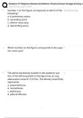
1. Number 1 on the figure corresponds to which of the left pulmonary artery
following?
a. lt pulmonary artery
b. ascending aorta
c. inferior vena cava
d. descending aorta
2. Which number on the figure corresponds to the supe- 4
rior vena cava?
3. The abnormal density located in the posterior por- D
tion of the left lung field on the figure has an avg
attenuation value of +5.0 HUs. This density most likely
represents:
a. pneumothorax
b. hemothorax
c. atelectasis
d. pleural effusion
, Mosby's CT Registry Review 3rd Edition: Practice Exam Images & Diagra
Study online at https://quizlet.com/_6p35i8
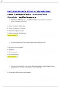
4. Number 2 on the figure corresponds to which of the left internal carotid artery
following?
a. left internal jugular vein
b. left external carotid artery
c. left internal carotid artery
d. left external jugular vein
5. Number 5 on the figure corresponds to which of the right internal jugular vein
following?
a. right internal jugular vein
b. right external carotid artery
c. right internal carotid artery
d. right external jugular vein
6. epiglottis
, Mosby's CT Registry Review 3rd Edition: Practice Exam Images & Diagra
Study online at https://quizlet.com/_6p35i8
Number 1 on the figure corresponds to which of the
following?
a. vocal cords
b. uvula
c. aryepiglottic fold
d. epiglottis
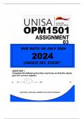
7. Number 1 on the figure corresponds to which of the stomach
following?
a. transverse colon
b. stomach
c. duodenum
d. hepatic flexure
8. Which of the following most likely describes the pt po- rt lat decub
sition during the formation of the image in the figure?
a. supine
b. prone
c. left lat decub
d. right lat decub
, Mosby's CT Registry Review 3rd Edition: Practice Exam Images & Diagra
Study online at https://quizlet.com/_6p35i8
9. CT exams of the abdomen are often performed in the duodenum and pancreat-
position on the figure to demonstrate the relationship ic head
between the:
a. ureters and renal collecting systems
b. duodenum and pancreatic head
c. large and small colon
d. liver and gallbladder
10. Number 3 on the figure corresponds to which of the right ureter
following?
a. right ureter
b. inferior vena cava
c. renal calculi
d. right renal artery
Study online at https://quizlet.com/_6p35i8
1. Number 1 on the figure corresponds to which of the left pulmonary artery
following?
a. lt pulmonary artery
b. ascending aorta
c. inferior vena cava
d. descending aorta
2. Which number on the figure corresponds to the supe- 4
rior vena cava?
3. The abnormal density located in the posterior por- D
tion of the left lung field on the figure has an avg
attenuation value of +5.0 HUs. This density most likely
represents:
a. pneumothorax
b. hemothorax
c. atelectasis
d. pleural effusion
, Mosby's CT Registry Review 3rd Edition: Practice Exam Images & Diagra
Study online at https://quizlet.com/_6p35i8
4. Number 2 on the figure corresponds to which of the left internal carotid artery
following?
a. left internal jugular vein
b. left external carotid artery
c. left internal carotid artery
d. left external jugular vein
5. Number 5 on the figure corresponds to which of the right internal jugular vein
following?
a. right internal jugular vein
b. right external carotid artery
c. right internal carotid artery
d. right external jugular vein
6. epiglottis
, Mosby's CT Registry Review 3rd Edition: Practice Exam Images & Diagra
Study online at https://quizlet.com/_6p35i8
Number 1 on the figure corresponds to which of the
following?
a. vocal cords
b. uvula
c. aryepiglottic fold
d. epiglottis
7. Number 1 on the figure corresponds to which of the stomach
following?
a. transverse colon
b. stomach
c. duodenum
d. hepatic flexure
8. Which of the following most likely describes the pt po- rt lat decub
sition during the formation of the image in the figure?
a. supine
b. prone
c. left lat decub
d. right lat decub
, Mosby's CT Registry Review 3rd Edition: Practice Exam Images & Diagra
Study online at https://quizlet.com/_6p35i8
9. CT exams of the abdomen are often performed in the duodenum and pancreat-
position on the figure to demonstrate the relationship ic head
between the:
a. ureters and renal collecting systems
b. duodenum and pancreatic head
c. large and small colon
d. liver and gallbladder
10. Number 3 on the figure corresponds to which of the right ureter
following?
a. right ureter
b. inferior vena cava
c. renal calculi
d. right renal artery