NR 603 APEA Predictor Exam Questions and
Answers Exam fully solved & updated 2025-
2026(latest version verified for accuracy) |
2024\2025Latest!!
Save
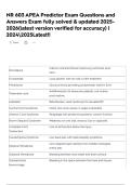
Calcium channel blocker improving outcomes post-
Nimodipine
SAH.
Furosemide Loop diuretic with no role in SAH treatment.
Prednisone Glucocorticoid providing symptomatic relief in SAH.
Antifibrinolytic for aneurysm patients, not routine
Tranexamic acid
post-rupture.
Labetalol Beta blocker, used cautiously for elevated BP.
Central Cord Syndrome Motor loss more severe in upper extremities.
Anterior Cord Syndrome Paraplegia with preserved posterior column function.
Brown-Séquard Syndrome Weakness on one side, sensory loss on opposite.
Complete Cord Loss of all function below the injury.
Transection
Cauda Equina Syndrome Low back pain with radiculopathy symptoms.
Lens-shaped hyper-density from middle meningeal
Epidural Hematoma
artery tear.
Subdural Hematoma Bleeding between dura mater and brain surface.
Subarachnoid Bleeding in the space between the brain and tissues.
Hemorrhage
, Intracerebral Hemorrhage Bleeding within the brain tissue itself.
Inflammation of protective membranes covering the
Meningitis
brain.
Hyperesthesia Increased sensitivity to sensory stimuli.
Mydriasis Dilated pupils, often indicating neurological issues.
Swelling of optic disc due to increased intracranial
Papilledema
pressure.
Neurological sign indicating upper motor neuron
Extensor Plantar Response
lesion.
Mass Effect Displacement of brain structures due to swelling.
Period of clarity after head injury before symptoms
Lucid Interval
worsen.
CT Scan Imaging technique used to diagnose brain injuries.
Traumatic Tear Injury causing a rupture in blood vessels.
Subdural hematoma Venous hemorrhage from ruptured bridging veins.
Epidural hematoma Arterial bleeding between skull and dura mater.
Subarachnoid Blood in subarachnoid space from aneurysm rupture.
hemorrhage
Blood accumulation in brain tissue due to
Intracerebral hemorrhage
hypertension.
Meningitis Inflammation of meninges, often with fever.
Lumbar puncture Diagnostic procedure to analyze cerebrospinal fluid.
Yellow discoloration in CSF indicating subarachnoid
Xanthochromia
hemorrhage.
Congenital aneurysm often causing subarachnoid
Berry aneurysm
hemorrhage.
Mass effect Pressure effect on brain structures from hematoma.
Contralateral hemiparesis Weakness on opposite side of brain injury.
Answers Exam fully solved & updated 2025-
2026(latest version verified for accuracy) |
2024\2025Latest!!
Save
Calcium channel blocker improving outcomes post-
Nimodipine
SAH.
Furosemide Loop diuretic with no role in SAH treatment.
Prednisone Glucocorticoid providing symptomatic relief in SAH.
Antifibrinolytic for aneurysm patients, not routine
Tranexamic acid
post-rupture.
Labetalol Beta blocker, used cautiously for elevated BP.
Central Cord Syndrome Motor loss more severe in upper extremities.
Anterior Cord Syndrome Paraplegia with preserved posterior column function.
Brown-Séquard Syndrome Weakness on one side, sensory loss on opposite.
Complete Cord Loss of all function below the injury.
Transection
Cauda Equina Syndrome Low back pain with radiculopathy symptoms.
Lens-shaped hyper-density from middle meningeal
Epidural Hematoma
artery tear.
Subdural Hematoma Bleeding between dura mater and brain surface.
Subarachnoid Bleeding in the space between the brain and tissues.
Hemorrhage
, Intracerebral Hemorrhage Bleeding within the brain tissue itself.
Inflammation of protective membranes covering the
Meningitis
brain.
Hyperesthesia Increased sensitivity to sensory stimuli.
Mydriasis Dilated pupils, often indicating neurological issues.
Swelling of optic disc due to increased intracranial
Papilledema
pressure.
Neurological sign indicating upper motor neuron
Extensor Plantar Response
lesion.
Mass Effect Displacement of brain structures due to swelling.
Period of clarity after head injury before symptoms
Lucid Interval
worsen.
CT Scan Imaging technique used to diagnose brain injuries.
Traumatic Tear Injury causing a rupture in blood vessels.
Subdural hematoma Venous hemorrhage from ruptured bridging veins.
Epidural hematoma Arterial bleeding between skull and dura mater.
Subarachnoid Blood in subarachnoid space from aneurysm rupture.
hemorrhage
Blood accumulation in brain tissue due to
Intracerebral hemorrhage
hypertension.
Meningitis Inflammation of meninges, often with fever.
Lumbar puncture Diagnostic procedure to analyze cerebrospinal fluid.
Yellow discoloration in CSF indicating subarachnoid
Xanthochromia
hemorrhage.
Congenital aneurysm often causing subarachnoid
Berry aneurysm
hemorrhage.
Mass effect Pressure effect on brain structures from hematoma.
Contralateral hemiparesis Weakness on opposite side of brain injury.