Acute rheumatic fever (ARF)
ARF is a systemic autoimmune disease triggered by cross-reactive immune responses between
group A β-hemolytic Streptococcus (GABHS) and host tissue epitopes, 2-4 weeks after infection.
Rheumatic fever is one of the main causes of cardiac surgery in children, mainly in low- and
middle-income countries. As GABHS can horizontally transfer genetic material, it is believed that
any GABHS can potentially acquire the ability to cause ARF. Genetic susceptibility is attributed to
several HLA-II alleles (ex. DR7, DR53) and polymorphisms in genes out of MHC gene (mannose-
binding lectin, TLR2).
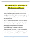
Bacterial immune evasion mechanisms
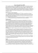
i) M-protein covers the bacterial cell wall. It’s a major virulence factor according to
which GABHS is differentiated into >240 M-types. The M-protein binds to the Fc portion of IgG
and IgA, creating an immunoglobulin shield with the Fab portion facing outward that prevents
phagocytosis and immune recognition.
ii) Within the M-protein lies a domain that binds to factor H, a complement regulatory
protein inhibiting antibody binding and complement-derived opsonin deposition.
Pathophysiology
ARF is associated with pharyngeal but not skin infection (for Aboriginals that’s not true).
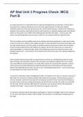
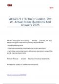
i) Humoral mechanisms (mimicry): the GAS M-protein bears a structural homology to
cardiac cytoskeletal tropomyosin and myosin and many other coiled structures (vimentin,
keratin, DNA, laminin). Epitope spreading (the process by which an immune response leads to
reaction against epitopes that are different from the original disease-triggering epitope) can
happen. The anti-myosin and anti-laminin IgG antibodies of patients with ARF bind to and
activate endothelial cells that express VCAM-1. Sydenham chorea is believed to be mediated by
antibodies that bind to neurons. IgG against streptococcal GlcNAc bind to antigens in the basal
ganglia and promote dopamine release. Arthritis in ARF is caused by deposit of immune
complexes.
ii) Cellular response: T cells (activated and expanded in the periphery) are attracted to
the heart by chemokines, cytokines and adhesion molecules. The autoreactive clones are
continuously activated by autoantigens. Both CD4+ and CD8+ invade the heart tissues, but the
CD4+ subset predominates and is the major effector of the inflammation that leads to valvular
damage. The peripheral and intracardiac T-cells of patients with ARF are also able to cross-
recognize several self-epitopes, chiefly cardiac myosin, laminin, tropomyosin and three specific
regions of the amino-terminal region of M protein. Recently, T cells producing IL-17/23 have
been described.
The initial and transient carditis, similar to chorea and arthritis, is believed to
be mediated primarily by humoral immunity (Th2 type), whereas severe and persistent carditis
is mediated by cellular immunity (Th1/17).
Clinical
Children tend to exhibit fever and carditis more frequently; arthritis is more common in adults.
The most common major manifestations during the first episode of ARF remain carditis, and
arthritis. Chorea (which have a female predominance), subcutaneous nodules (<10%) and
erythema marginatum (<6%) remain much less common but highly specific manifestations of.
Cardiac involvement (50-70%): loosely classified as acute and chronic. The carditis
associated with ARF is classically considered to be a pancarditis. Congestive heart failure (5-10%)
, is the most life-threatening feature of ARF and can develop if severe valvular damage occurs in
addition to cardiac dysfunction.
-Valvulitis: the predominant manifestation of carditis, especially of the mitral
and aortic valves (MV > AoV). Mitral regurgitation is the most common early valvular finding and
may be accompanied by aortic regurgitation.
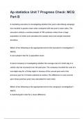
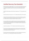
-Myocarditis: initially nonspecific myocarditis, later granulomatous myocarditis
with Aschoff nodules beneath the endocardium close to or at the valve. Aschoff nodules are
composed of a central area of fibrinoid change that is surrounded by lymphocytic infiltration
(the majority CD4+ and occasionally CD8+ and plasma cells), specialized histiocytes that are
referred to as Aschoff giant (multinucleated) cells and Anitschkow cells (“caterpillar cells,” due
to the appearance of its chromatin). Myocarditis in the absence of valvulitis is unlikely to be
ARF.
-Endocarditis: in early stages, it is associated with edema of the valves and
possibly vegetations along the free borders of the cusps. These minute vegetations are platelet-
rich microthrombi (marantic vegetations), which do not become dislodged and hence do not
produce emboli. When later fibrosis occurs, it usually doesn’t affect the function of the valve.
However, in others, the scarring is progressive over years and may affect the valves leading to
contraction of the cuspid or thickening and stiffening of the cusp, leading to valvular
incompetence or the fusion of the commissures, resulting in stenosis.
-Pericarditis: fibrin-rich exudate, which leaves fibrosis and adhesions.
Chronic rheumatic heart disease: the single greatest cause of acquired
valvular disease in the world, responsible for 25% heart failure cases in endemic countries. In
the majority of cases, the valvular disease occurs 10-20yrs after the original attack. Patients
<30yrs generally present pure mitral regurgitation, whereas middle-aged adults develop mitral
stenosis. MV involvement is nearly universal; the AoV is involved in 20-30%.
Arthritis (60-80%): migratory, in an additive/remitting pattern and the affected sites
generally overlap in time. The earliest sign and lasts for a few days to a week for each joint,
however, the entire articular picture is intense for 1 week and may linger for another 1-2 weeks.
If joint swelling persists >4wks, it is necessary to reconsider the diagnosis. The knees, ankles,
elbows and wrists are most commonly affected. Repetitive attacks can lead to Jaccoud
arthropathy. Usually these joint manifestations are self-limiting and subside with or without
treatment by 4 weeks. Polyarthralgia and aseptic monoarthritis are commonly seen in endemic
regions and are considered major manifestations for diagnosis in moderate-to-high-risk
populations in the 2015 Jones criteria, while monoarthralgia is considered a minor
manifestation.
Chorea (10–30%): is considered a major manifestation. Evidence of preceding
streptococcal infection can be absent because of the long delay to manifestation. Cardiac
involvement is common in patients with chorea. Up to 90% have carditis when subclinical
involvement is also considered, that’s why a diagnosis of chorea should be accompanied by a
heart u/s.
Skin (<10%): skin manifestations are uncommon but specific for ARF and are major
criteria for diagnosis.
-Subcutaneous nodules (<10%): smaller and last for a shorter period than those
of RA, usually located over a bony surface or prominence or near tendons (usually extensor
surfaces) and usually symmetric. The elbows are involved most frequently. More common in
patients with a prolonged history of active carditis than those in the early stages of the disease.
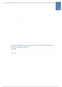
-Erythema marginatum (annular erythema) (<6%) is infrequent in ARF, affecting
the limbs and trunk but spares the face. It is evanescent, pink, non-pruritic and non-tender that
ARF is a systemic autoimmune disease triggered by cross-reactive immune responses between
group A β-hemolytic Streptococcus (GABHS) and host tissue epitopes, 2-4 weeks after infection.
Rheumatic fever is one of the main causes of cardiac surgery in children, mainly in low- and
middle-income countries. As GABHS can horizontally transfer genetic material, it is believed that
any GABHS can potentially acquire the ability to cause ARF. Genetic susceptibility is attributed to
several HLA-II alleles (ex. DR7, DR53) and polymorphisms in genes out of MHC gene (mannose-
binding lectin, TLR2).
Bacterial immune evasion mechanisms
i) M-protein covers the bacterial cell wall. It’s a major virulence factor according to
which GABHS is differentiated into >240 M-types. The M-protein binds to the Fc portion of IgG
and IgA, creating an immunoglobulin shield with the Fab portion facing outward that prevents
phagocytosis and immune recognition.
ii) Within the M-protein lies a domain that binds to factor H, a complement regulatory
protein inhibiting antibody binding and complement-derived opsonin deposition.
Pathophysiology
ARF is associated with pharyngeal but not skin infection (for Aboriginals that’s not true).
i) Humoral mechanisms (mimicry): the GAS M-protein bears a structural homology to
cardiac cytoskeletal tropomyosin and myosin and many other coiled structures (vimentin,
keratin, DNA, laminin). Epitope spreading (the process by which an immune response leads to
reaction against epitopes that are different from the original disease-triggering epitope) can
happen. The anti-myosin and anti-laminin IgG antibodies of patients with ARF bind to and
activate endothelial cells that express VCAM-1. Sydenham chorea is believed to be mediated by
antibodies that bind to neurons. IgG against streptococcal GlcNAc bind to antigens in the basal
ganglia and promote dopamine release. Arthritis in ARF is caused by deposit of immune
complexes.
ii) Cellular response: T cells (activated and expanded in the periphery) are attracted to
the heart by chemokines, cytokines and adhesion molecules. The autoreactive clones are
continuously activated by autoantigens. Both CD4+ and CD8+ invade the heart tissues, but the
CD4+ subset predominates and is the major effector of the inflammation that leads to valvular
damage. The peripheral and intracardiac T-cells of patients with ARF are also able to cross-
recognize several self-epitopes, chiefly cardiac myosin, laminin, tropomyosin and three specific
regions of the amino-terminal region of M protein. Recently, T cells producing IL-17/23 have
been described.
The initial and transient carditis, similar to chorea and arthritis, is believed to
be mediated primarily by humoral immunity (Th2 type), whereas severe and persistent carditis
is mediated by cellular immunity (Th1/17).
Clinical
Children tend to exhibit fever and carditis more frequently; arthritis is more common in adults.
The most common major manifestations during the first episode of ARF remain carditis, and
arthritis. Chorea (which have a female predominance), subcutaneous nodules (<10%) and
erythema marginatum (<6%) remain much less common but highly specific manifestations of.
Cardiac involvement (50-70%): loosely classified as acute and chronic. The carditis
associated with ARF is classically considered to be a pancarditis. Congestive heart failure (5-10%)
, is the most life-threatening feature of ARF and can develop if severe valvular damage occurs in
addition to cardiac dysfunction.
-Valvulitis: the predominant manifestation of carditis, especially of the mitral
and aortic valves (MV > AoV). Mitral regurgitation is the most common early valvular finding and
may be accompanied by aortic regurgitation.
-Myocarditis: initially nonspecific myocarditis, later granulomatous myocarditis
with Aschoff nodules beneath the endocardium close to or at the valve. Aschoff nodules are
composed of a central area of fibrinoid change that is surrounded by lymphocytic infiltration
(the majority CD4+ and occasionally CD8+ and plasma cells), specialized histiocytes that are
referred to as Aschoff giant (multinucleated) cells and Anitschkow cells (“caterpillar cells,” due
to the appearance of its chromatin). Myocarditis in the absence of valvulitis is unlikely to be
ARF.
-Endocarditis: in early stages, it is associated with edema of the valves and
possibly vegetations along the free borders of the cusps. These minute vegetations are platelet-
rich microthrombi (marantic vegetations), which do not become dislodged and hence do not
produce emboli. When later fibrosis occurs, it usually doesn’t affect the function of the valve.
However, in others, the scarring is progressive over years and may affect the valves leading to
contraction of the cuspid or thickening and stiffening of the cusp, leading to valvular
incompetence or the fusion of the commissures, resulting in stenosis.
-Pericarditis: fibrin-rich exudate, which leaves fibrosis and adhesions.
Chronic rheumatic heart disease: the single greatest cause of acquired
valvular disease in the world, responsible for 25% heart failure cases in endemic countries. In
the majority of cases, the valvular disease occurs 10-20yrs after the original attack. Patients
<30yrs generally present pure mitral regurgitation, whereas middle-aged adults develop mitral
stenosis. MV involvement is nearly universal; the AoV is involved in 20-30%.
Arthritis (60-80%): migratory, in an additive/remitting pattern and the affected sites
generally overlap in time. The earliest sign and lasts for a few days to a week for each joint,
however, the entire articular picture is intense for 1 week and may linger for another 1-2 weeks.
If joint swelling persists >4wks, it is necessary to reconsider the diagnosis. The knees, ankles,
elbows and wrists are most commonly affected. Repetitive attacks can lead to Jaccoud
arthropathy. Usually these joint manifestations are self-limiting and subside with or without
treatment by 4 weeks. Polyarthralgia and aseptic monoarthritis are commonly seen in endemic
regions and are considered major manifestations for diagnosis in moderate-to-high-risk
populations in the 2015 Jones criteria, while monoarthralgia is considered a minor
manifestation.
Chorea (10–30%): is considered a major manifestation. Evidence of preceding
streptococcal infection can be absent because of the long delay to manifestation. Cardiac
involvement is common in patients with chorea. Up to 90% have carditis when subclinical
involvement is also considered, that’s why a diagnosis of chorea should be accompanied by a
heart u/s.
Skin (<10%): skin manifestations are uncommon but specific for ARF and are major
criteria for diagnosis.
-Subcutaneous nodules (<10%): smaller and last for a shorter period than those
of RA, usually located over a bony surface or prominence or near tendons (usually extensor
surfaces) and usually symmetric. The elbows are involved most frequently. More common in
patients with a prolonged history of active carditis than those in the early stages of the disease.
-Erythema marginatum (annular erythema) (<6%) is infrequent in ARF, affecting
the limbs and trunk but spares the face. It is evanescent, pink, non-pruritic and non-tender that