Lecture 1: membrane potential
Equilibrium potential:
Each ion has its own equilibrium potential. Sodium and chloride are higher outside and potassium is
higher inside. These gradients are maintained by ion transporters.
1. Assume membrane potential is 0mV
2. Asymmetric K+ causes K+ efflux. Potassium goes out, because there are less potassium
ions on the outside of the cell (chemical driving force)
3. Membrane potential becomes negative. There comes an electrical driving force inside
the cell, because the cell gets more negative on the inside.
4. K+ efflux still continues
5. Membrane potential becomes more negative
6. No more K+ efflux
=> equilibrium potential: electrical driving force = chemical driving force.
You can calculate the equilibrium with the Nernst equation.
Ion channels have an effect on the resting membrane potential
and the membrane potential in response to synaptic inputs (signals). If an ion channel opens, the
membrane potential shift towards the equiluibrium potential of the ion. The equiluibrium potential os
ion X is the Vm (membrane potential) when there is no net current is flowing when channels are open.
Each ion has its own equiluibrium potential (E k, Ena, Ecl). The reversal potential of AMPA receptors is 0
mV, because the permeability for potassium and sodium ions is rougly equal.
(Resting) membrane potential:
The resting membrane potential of a neuron is about -60/-70 mV. The Goldman Hodgkin Katz
equation says that the resting membrane potential is a combination of equilibrium of all ions. The
impact of individual ion channels is determined by their permeability (ion conductance). Because
many potassium channels are open at rest (higher permeability): V rest is closer to the equiluibrium
potential of potassium than sodium. The P is the permeability factor (conductance), the C are ion
concentrations.
Action potential:
Sodium channels have fast kinetics (open and close rapidly),
potassium channels have slower kinetics.
1. Assume membrane potential is at rest (-60mV)
2. Stimulation depolarizes membrane potential (the driving force is a
little bit less).
3. Na+ channels open (because of the voltage change)
4. Na+ influx
5. More depolarization
6. More opening of Na+ channels (positive feedback)
7. K+ channel opens & Na+ inactivation -> beginning of the refractory period (negative
feedback
8. K+ efflux (no more Na+ influx)
9. Hyperpolarization
10. Na+ channels close
11. K+ channels close
12. Na+ channel inactivation is removed
,An action potential is initiated at the axon hillock and propagates toward the axon terminals. When
there are two action potentials generated, they collide and they cannot propagate further. This is
because an action potential is followed by a refractory period. Myelin speeds up the conduction
velocity.
Ion pump (transporter) Ion channel
Actively move selected ions against Allow ions to diffuse down concentration
concentration gradient gradient
Create ion concentration gradients Are selectively permeable to certain ions
Maintains concentration differences Change membrane permeability
Important for resting conditions Produces signals (action)
Protein structure of ion channels:
Ion channels are just protein structures on the membrane. A potassium channel has four sub units.
There are voltage-dependent channels and ligand-dependent channels (glutamate receptors/AMPA
receptor) they open when glutamate/calcium binds. Ion channels can be sensitive to voltage,
neurotransmitter concentration, pH, temperature, mechanical stress, light etc.
Channel rhodopsin are ion channels that open based on light. With coloured light you can open or
close the ion channel -> remote control, specific expression is possible. Voltage clamp method.
Lecture 2: Neuron types
Types of neurons – excitatory vs inhibitory neurons:
80/90% of the neurons are excitatory and 10/20% are inhibitory. Excitatory neurons make excitatory
synaptic contact to postsynaptic cells and inhibitory neurons make inhibitory synaptic contacts. All
neurons receive both excitatory (glutamatergic) and inhibitory (GABAergic) synapses. An excitatory
neuron makes a negative synaptic signal (EPSP) and an inhibitory neuron makes a positive synaptic
signal (IPSP). There is a healthy balance between excitation and inhibition.
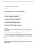
The neocortex is the outermost part of the cerebrum
and has six layers. Every layer has their own kind of
neurons. -> excitatory neurons. Layer I is not involved,
because there are only inhibitory neurons. Layer II/III
and V make connections with layer I (a lot of
modulation). There are pyramidal cells in layer II, III,
IV, V and VI. they have an apical dendrite and a
pyramidal shaped soma. A spiny stellate cell is in layer
IV and have no apical dendrite and are
multipolar.
<- inhibitory neurons.
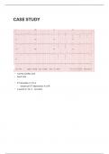
The hippocampus is an important brain area,
because it’s very plastic. There can be made
changes in your hippocampal connectivity. It is
a short-term memory place.
, In the hippocampus, pyramidal cells are densely packed in one layer (pyramidal layer/CA1) they have
their somata in pyramidal layer (SP) and pyramidal cells on the dorsal part are upside down (apical
dendrites go downwards). Inhitory neurons have a wide variety of morphologies. Inhibitory cells are
also called interneurons and pyramidal cells are called principal cells.
Classification of inhibitory neurons:
There are three main approaches:
- Morphology (shapes, location, synaptic projections)
o The cell body (soma) can has different shapes and orientations.
o The neural processes (dendrites and axons).
Unipolar neurons have a single primary process. One branch serves as the
axon, other branches function as receiving structures. They are dominant in
the nervous systems of invertebrates, in vertebrates in the ANS.
Bipolar neurons have a soma that gives rise to a dendrite and an axon. Many
sensory cells, in the retina and olfactory epithelium of the nose.
Pseudo-unipolar cells are receptor neurons that convey touch, pressure and
pain signals to the spinal cord.
Multipolar neurons predominate in the nervous system of vertebrates.
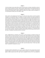
- Electrophysiological properties (firing patterns, shapes of action potentials and ion channels)
o Responses to both negative and positive current injections are checked.
o This depolarizing voltage sag is caused by an inward Ih current via hyperpolarization-
activated cyclic nucleotide-gated channels (permeable for sodium and potassium). If
you put an Ih blocker, the sag is gone because the current is going up again.
o Firing patterns: different sodium and potassium channel subtypes.
Regular spiking: not capable of showing high frequency spiking, even with a
large current injection.
Fast spiking: capable of showing high frequency spiking.
Intrinsically bursting: shows trains of high frequency spiking for a short
period. After the initial burst, AP firing is highly regular.
Chattering
Adapting firing: inter-spike interval increases in time. The interval between
Aps increases over time.
- Molecular properties (biomarkers, gene expression, proteins and peptides)
o Co-labelling of PV+ (parvalbumin) and SST+ (somatostatin) cells in hippocampus. PV is
a calcium binding protein, somatostatin is a neuropeptide and vasoactive intestinal
peptide (VIP) also.
o PV cells are also called basket cells. They have axonal projection to perisomatic region
and have fast spiking firing pattern.
o Axo-axonic cells (chandelier cells) are also PV positive, have a fast spiking firing
pattern and axonal projection to initial segment of axons. These are even more
powerful.
o Distal dendrite targeting cells are mostly SST+. Axonal projection to distal dendrite
and have a regular spiking firing pattern. They are less powerful, but more precise.
o Interneuron-targeting cells. Interneurons that target other interneurons. They can
project to the dendrites and somas. VIP positive and have a regular spiking firing
pattern.
Equilibrium potential:
Each ion has its own equilibrium potential. Sodium and chloride are higher outside and potassium is
higher inside. These gradients are maintained by ion transporters.
1. Assume membrane potential is 0mV
2. Asymmetric K+ causes K+ efflux. Potassium goes out, because there are less potassium
ions on the outside of the cell (chemical driving force)
3. Membrane potential becomes negative. There comes an electrical driving force inside
the cell, because the cell gets more negative on the inside.
4. K+ efflux still continues
5. Membrane potential becomes more negative
6. No more K+ efflux
=> equilibrium potential: electrical driving force = chemical driving force.
You can calculate the equilibrium with the Nernst equation.
Ion channels have an effect on the resting membrane potential
and the membrane potential in response to synaptic inputs (signals). If an ion channel opens, the
membrane potential shift towards the equiluibrium potential of the ion. The equiluibrium potential os
ion X is the Vm (membrane potential) when there is no net current is flowing when channels are open.
Each ion has its own equiluibrium potential (E k, Ena, Ecl). The reversal potential of AMPA receptors is 0
mV, because the permeability for potassium and sodium ions is rougly equal.
(Resting) membrane potential:
The resting membrane potential of a neuron is about -60/-70 mV. The Goldman Hodgkin Katz
equation says that the resting membrane potential is a combination of equilibrium of all ions. The
impact of individual ion channels is determined by their permeability (ion conductance). Because
many potassium channels are open at rest (higher permeability): V rest is closer to the equiluibrium
potential of potassium than sodium. The P is the permeability factor (conductance), the C are ion
concentrations.
Action potential:
Sodium channels have fast kinetics (open and close rapidly),
potassium channels have slower kinetics.
1. Assume membrane potential is at rest (-60mV)
2. Stimulation depolarizes membrane potential (the driving force is a
little bit less).
3. Na+ channels open (because of the voltage change)
4. Na+ influx
5. More depolarization
6. More opening of Na+ channels (positive feedback)
7. K+ channel opens & Na+ inactivation -> beginning of the refractory period (negative
feedback
8. K+ efflux (no more Na+ influx)
9. Hyperpolarization
10. Na+ channels close
11. K+ channels close
12. Na+ channel inactivation is removed
,An action potential is initiated at the axon hillock and propagates toward the axon terminals. When
there are two action potentials generated, they collide and they cannot propagate further. This is
because an action potential is followed by a refractory period. Myelin speeds up the conduction
velocity.
Ion pump (transporter) Ion channel
Actively move selected ions against Allow ions to diffuse down concentration
concentration gradient gradient
Create ion concentration gradients Are selectively permeable to certain ions
Maintains concentration differences Change membrane permeability
Important for resting conditions Produces signals (action)
Protein structure of ion channels:
Ion channels are just protein structures on the membrane. A potassium channel has four sub units.
There are voltage-dependent channels and ligand-dependent channels (glutamate receptors/AMPA
receptor) they open when glutamate/calcium binds. Ion channels can be sensitive to voltage,
neurotransmitter concentration, pH, temperature, mechanical stress, light etc.
Channel rhodopsin are ion channels that open based on light. With coloured light you can open or
close the ion channel -> remote control, specific expression is possible. Voltage clamp method.
Lecture 2: Neuron types
Types of neurons – excitatory vs inhibitory neurons:
80/90% of the neurons are excitatory and 10/20% are inhibitory. Excitatory neurons make excitatory
synaptic contact to postsynaptic cells and inhibitory neurons make inhibitory synaptic contacts. All
neurons receive both excitatory (glutamatergic) and inhibitory (GABAergic) synapses. An excitatory
neuron makes a negative synaptic signal (EPSP) and an inhibitory neuron makes a positive synaptic
signal (IPSP). There is a healthy balance between excitation and inhibition.
The neocortex is the outermost part of the cerebrum
and has six layers. Every layer has their own kind of
neurons. -> excitatory neurons. Layer I is not involved,
because there are only inhibitory neurons. Layer II/III
and V make connections with layer I (a lot of
modulation). There are pyramidal cells in layer II, III,
IV, V and VI. they have an apical dendrite and a
pyramidal shaped soma. A spiny stellate cell is in layer
IV and have no apical dendrite and are
multipolar.
<- inhibitory neurons.
The hippocampus is an important brain area,
because it’s very plastic. There can be made
changes in your hippocampal connectivity. It is
a short-term memory place.
, In the hippocampus, pyramidal cells are densely packed in one layer (pyramidal layer/CA1) they have
their somata in pyramidal layer (SP) and pyramidal cells on the dorsal part are upside down (apical
dendrites go downwards). Inhitory neurons have a wide variety of morphologies. Inhibitory cells are
also called interneurons and pyramidal cells are called principal cells.
Classification of inhibitory neurons:
There are three main approaches:
- Morphology (shapes, location, synaptic projections)
o The cell body (soma) can has different shapes and orientations.
o The neural processes (dendrites and axons).
Unipolar neurons have a single primary process. One branch serves as the
axon, other branches function as receiving structures. They are dominant in
the nervous systems of invertebrates, in vertebrates in the ANS.
Bipolar neurons have a soma that gives rise to a dendrite and an axon. Many
sensory cells, in the retina and olfactory epithelium of the nose.
Pseudo-unipolar cells are receptor neurons that convey touch, pressure and
pain signals to the spinal cord.
Multipolar neurons predominate in the nervous system of vertebrates.
- Electrophysiological properties (firing patterns, shapes of action potentials and ion channels)
o Responses to both negative and positive current injections are checked.
o This depolarizing voltage sag is caused by an inward Ih current via hyperpolarization-
activated cyclic nucleotide-gated channels (permeable for sodium and potassium). If
you put an Ih blocker, the sag is gone because the current is going up again.
o Firing patterns: different sodium and potassium channel subtypes.
Regular spiking: not capable of showing high frequency spiking, even with a
large current injection.
Fast spiking: capable of showing high frequency spiking.
Intrinsically bursting: shows trains of high frequency spiking for a short
period. After the initial burst, AP firing is highly regular.
Chattering
Adapting firing: inter-spike interval increases in time. The interval between
Aps increases over time.
- Molecular properties (biomarkers, gene expression, proteins and peptides)
o Co-labelling of PV+ (parvalbumin) and SST+ (somatostatin) cells in hippocampus. PV is
a calcium binding protein, somatostatin is a neuropeptide and vasoactive intestinal
peptide (VIP) also.
o PV cells are also called basket cells. They have axonal projection to perisomatic region
and have fast spiking firing pattern.
o Axo-axonic cells (chandelier cells) are also PV positive, have a fast spiking firing
pattern and axonal projection to initial segment of axons. These are even more
powerful.
o Distal dendrite targeting cells are mostly SST+. Axonal projection to distal dendrite
and have a regular spiking firing pattern. They are less powerful, but more precise.
o Interneuron-targeting cells. Interneurons that target other interneurons. They can
project to the dendrites and somas. VIP positive and have a regular spiking firing
pattern.