General Anatomical Overview – GI tract
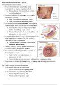
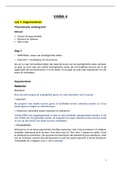
The digestive tract exists as follows:
1. Food is immediately taken up by the oral cavity
responsible for the physical breakdown of food.
a. Salivary Glands: The submandibular, parotid,
and sublingual glands
2. Swallowed food enters the esophagus, connecting the
stomach and oral cavity
a. Upper ⅓ propels food using skeletal muscle
b. Lower ⅔ propels food using smooth muscle
3. The esophagus connects the the stomach, responsible for
continued digest, mixing food with acid and creating chyme
a. The stomach is composed of three regions: Upper
fundus, Central body, Lower antrum
b. The stomach connects to the S.I. via the pylorus and
pyloric valve (smooth muscle)
4. Integrated signals regulate the rate at which chyme enters the
small intestine via the pylorus. The small intestine is
responsible for most of the digestion.
a. The small intestine is composed of three regions:
duodenum, jejunum, ileum.
5. Digestion in the SI is aided by intestinal enzymes and
exocrine secretion by the pancreas and liver.
6. Food continues into the large intestine, responsible for
the absorption of nutrients
a. Colon (1st ½): water and electrolytes are
absorbed into the ECF
b. Rectum (terminal section): distension of wall responsible for defecation reflex.
7. Feces leaves the GI tract via the anus through the external anal sphincter (skeletal muscle)
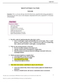
The GI wall is crumpled to increase surface area
- In the stomach these folds are called rugae
- Possess tubular invagination: gastric glands
- In the small intestine these folds are called plicae
- Possess fingerlike projections: villi
- Possess tubular invaginations: crypts
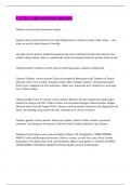
,Layers of GI tract Wall
The gut wall is composed of four layers:
- Inner mucosa: inner layer of epithelium facing the lumen.
- Submucosa:
- Muscularis Externa: layers of smooth muscle (2).
- Serosa: outermost layer of connective tissue.
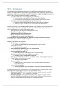
#1 – Inner Mucosa: Epithelial layer of GI tract.
The inner mucosa is composed of three layers:
1. Mucosal epithelium: epithelial layer of GI tract
a. Contains transporting epithelial cells, endocrine and exocrine secretory cells, stem cells.
b. At the mucosal surface: cells secreted ions, enzymes, mucus, and paracrine molecules.
c. At the serosal surface: substances absorbed pass into the ECF
d. Cell-to-cell junctions are tight in the stomach and jejunum, but
e. GI stem cells are rapidly dividing. Newly formed cells move from bottom of crypt to top
2. Lamina Propria: subepithelial connective tissue
a. Contains nerve fibers and blood vessels to supply epithelium
b. Contains wandering immune cells
c. Collections of lymphoid tissue adjoining the epithelium form small nodules and larger Peyer’s
Patches, a major part of gut-associated lymphoid tissue
3. Muscularis Mucosae: thin layer of smooth muscle
a. Contraction of muscle in this layer alters effective surface area
#2 – Submucosa: Connective tissue layer of GI tract
- Composed of connective tissue with larger blood and lymph vessels
- Contains submucosal plexus (one of two major nerve networks of the enteric NS)
, - Innervates both epithelial layer and muscularis mucosae
#3 – Muscularis Externa: Outer wall of GI tract, consists of two layers of smooth muscle
- Inner smooth muscle: circular → decreases diameter of tube
- Outer smooth muscle: Longitudinal → shortens length of tube
- Contains myenteric plexus (second of two major nerve networks in enteric NS)
- Innervates the muscular activity of the muscularis externa
#4 – Serosa: Continuation of the peritoneal membrane, covering the GI tract.
- Peritoneum also forms mesentery
Digestion Function & processes:
The primary function of the digestive system is to move nutrients, water, and electrolytes from the external
environment into the body’s external environment. This is accomplished by:
1. Digestion: the chemical and mechanical breakdown of foods.
2. Absorption: the movement of substances from the lumen into the ECF.
3. Secretion: The movement of substances from the ECF to the lumen OR the release of substances
synthesized by GI epithelial cells into the lumen or ECF.
4. Motility: The movement of material in the GI tract, as a result of muscular contraction.
- Motility and secretion are the most regulated.
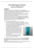
The GI tract faces three significant challenges.
1. Avoiding autodigestion: Enzymes used to digest food cannot digest host cells. When protective
mechanisms fail → peptic ulcers.
2. Mass balance: Must match fluid input with output
a. Input of fluid = 2L/day, exocrine/endocrine output = 7L/day
3. Vomiting and diarrhea
4. Immune Defense
Secretion
Each day, we secrete more fluids than we ingest. Secretions include:
1. Digestive Enzymes: Secreted either by exocrine glands (pancreas and salivary glands) or epithelial
cells (stomach and S.I.)
a. Peptide hormones: synthesized on RER, packaged by GA, released on demand by exocytosis.
b. Secreted inactively so as to not damage cells known as zymogens (e.g. pepsinogen)
2. Mucus: Secretion of glycoproteins called mucins.
a. Lubricate the contents of the gut and protect the gut
b. Secreted by mucous cells in the stomach and goblet cells in the intestine
c. Parasympathetic innervation triggers release
The digestive tract exists as follows:
1. Food is immediately taken up by the oral cavity
responsible for the physical breakdown of food.
a. Salivary Glands: The submandibular, parotid,
and sublingual glands
2. Swallowed food enters the esophagus, connecting the
stomach and oral cavity
a. Upper ⅓ propels food using skeletal muscle
b. Lower ⅔ propels food using smooth muscle
3. The esophagus connects the the stomach, responsible for
continued digest, mixing food with acid and creating chyme
a. The stomach is composed of three regions: Upper
fundus, Central body, Lower antrum
b. The stomach connects to the S.I. via the pylorus and
pyloric valve (smooth muscle)
4. Integrated signals regulate the rate at which chyme enters the
small intestine via the pylorus. The small intestine is
responsible for most of the digestion.
a. The small intestine is composed of three regions:
duodenum, jejunum, ileum.
5. Digestion in the SI is aided by intestinal enzymes and
exocrine secretion by the pancreas and liver.
6. Food continues into the large intestine, responsible for
the absorption of nutrients
a. Colon (1st ½): water and electrolytes are
absorbed into the ECF
b. Rectum (terminal section): distension of wall responsible for defecation reflex.
7. Feces leaves the GI tract via the anus through the external anal sphincter (skeletal muscle)
The GI wall is crumpled to increase surface area
- In the stomach these folds are called rugae
- Possess tubular invagination: gastric glands
- In the small intestine these folds are called plicae
- Possess fingerlike projections: villi
- Possess tubular invaginations: crypts
,Layers of GI tract Wall
The gut wall is composed of four layers:
- Inner mucosa: inner layer of epithelium facing the lumen.
- Submucosa:
- Muscularis Externa: layers of smooth muscle (2).
- Serosa: outermost layer of connective tissue.
#1 – Inner Mucosa: Epithelial layer of GI tract.
The inner mucosa is composed of three layers:
1. Mucosal epithelium: epithelial layer of GI tract
a. Contains transporting epithelial cells, endocrine and exocrine secretory cells, stem cells.
b. At the mucosal surface: cells secreted ions, enzymes, mucus, and paracrine molecules.
c. At the serosal surface: substances absorbed pass into the ECF
d. Cell-to-cell junctions are tight in the stomach and jejunum, but
e. GI stem cells are rapidly dividing. Newly formed cells move from bottom of crypt to top
2. Lamina Propria: subepithelial connective tissue
a. Contains nerve fibers and blood vessels to supply epithelium
b. Contains wandering immune cells
c. Collections of lymphoid tissue adjoining the epithelium form small nodules and larger Peyer’s
Patches, a major part of gut-associated lymphoid tissue
3. Muscularis Mucosae: thin layer of smooth muscle
a. Contraction of muscle in this layer alters effective surface area
#2 – Submucosa: Connective tissue layer of GI tract
- Composed of connective tissue with larger blood and lymph vessels
- Contains submucosal plexus (one of two major nerve networks of the enteric NS)
, - Innervates both epithelial layer and muscularis mucosae
#3 – Muscularis Externa: Outer wall of GI tract, consists of two layers of smooth muscle
- Inner smooth muscle: circular → decreases diameter of tube
- Outer smooth muscle: Longitudinal → shortens length of tube
- Contains myenteric plexus (second of two major nerve networks in enteric NS)
- Innervates the muscular activity of the muscularis externa
#4 – Serosa: Continuation of the peritoneal membrane, covering the GI tract.
- Peritoneum also forms mesentery
Digestion Function & processes:
The primary function of the digestive system is to move nutrients, water, and electrolytes from the external
environment into the body’s external environment. This is accomplished by:
1. Digestion: the chemical and mechanical breakdown of foods.
2. Absorption: the movement of substances from the lumen into the ECF.
3. Secretion: The movement of substances from the ECF to the lumen OR the release of substances
synthesized by GI epithelial cells into the lumen or ECF.
4. Motility: The movement of material in the GI tract, as a result of muscular contraction.
- Motility and secretion are the most regulated.
The GI tract faces three significant challenges.
1. Avoiding autodigestion: Enzymes used to digest food cannot digest host cells. When protective
mechanisms fail → peptic ulcers.
2. Mass balance: Must match fluid input with output
a. Input of fluid = 2L/day, exocrine/endocrine output = 7L/day
3. Vomiting and diarrhea
4. Immune Defense
Secretion
Each day, we secrete more fluids than we ingest. Secretions include:
1. Digestive Enzymes: Secreted either by exocrine glands (pancreas and salivary glands) or epithelial
cells (stomach and S.I.)
a. Peptide hormones: synthesized on RER, packaged by GA, released on demand by exocytosis.
b. Secreted inactively so as to not damage cells known as zymogens (e.g. pepsinogen)
2. Mucus: Secretion of glycoproteins called mucins.
a. Lubricate the contents of the gut and protect the gut
b. Secreted by mucous cells in the stomach and goblet cells in the intestine
c. Parasympathetic innervation triggers release