· helm Q+A
Sh review
⑨ other
1UNIT 6
CHAPTER 27
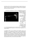
CYSTIC KIDNEY DISEASE
● Genetically transmitted renal disorder resulting in fluid-filled (localized to 1 or affect both
f kidneys)
● Common in men & increase w/ aging
● Can lead to RF, requiring dialysis or transplant
Imonitor
failure)
● 2 types:
river ○ Autosomal recessive forms polycystic kidney disease
■ Dx. in infants & young children
○ Autosomal dominant types polycystic kidney disease
■ May not be apparent until adulthood
POLYCYSTIC KIDNEY DISEASE ↳ get genetic testing
● Genetic component (parents pass to children)
● Have pt see genetic counselor
AUTOSOMAL RECESSIVE POLYCYSTIC KIDNEY
● Patho/etiology:
○ Identified in neonatal period *at birth*
○ When accompanied by pulmonary hypoplasia (lungs not developed) may result in
death
○ If survives to adulthood, retain some RF but will decline in liver function
● Manifestations:
○ Resp distress
○ Palpable kidneys
○ Systemic HTN
● DX: Ultrasound, CT, MRI
AUTOSOMAL DOMINANT POLYCYSTIC KIDNEY
● Patho/etiology:
○ Most common hereditary cystic KD involving both kidneys
○ Can occur at diff rates
monitor
-
○ Eventually results in end stage KD
O
○ Cysts can multiply & expand, kidney size ↑ & ↓ seen in GFR
○ Cysts can move to other organs → most common *Liver*
○ 2 Types:
■ Chromosome 16 - PKD1
■ Chromosome 4 - PKD2
● Manifestations:
○ Early middle-middle adulthood (40-59 y/o)
○ Ability to concentrate urine is ↓
-
○ HTN, proteinuria, hematuria
○ Pain from bleeding
8
○ Mvt of kidney stones
○ UTIs
○ Adults: PAIN is a freq complaint
● DX: Family hx, LIVER biopsy, imaging
Labs to know:
Albumin: 3.5-5 HCT: Male: 42-52% Female: 37-47%
BUN: 10-20 GFR: 90-120mL/min
Creatinine: Male: 0.6-1.2 Female: 0.5-1.1 RBC: Male: 4.7-6.1 Female: 4.2-5.4
Potassium: 3.5-5 *Triglycerides: Male: 40-160 Female: 35-135
HGB: Male: 14-18 Female: 12-16
, * *
ACUTE PYELONEPHRITIS CHRONIC PYELONEPHRITIS
● Infect of the renal pelvis & kidney tubules d/t Atrophied kidneys w/ diffuse scarring
ascending UTI
● Kidney infect: when UTI goes to the kidneys Patho/etiology
● Chronic reflux of urine into renal pelvis
Patho/etiology
● Can result in CKD G
⑧● Obstruction/ureteral reflux that allows RF:
contaminated urine to enter kidneys
⑨
● Renal calculi, neurogenic bladder,
● Highest incidence: infants, women & elderly w/ vesicoureteral reflux, intrarenal disease
Because
E.Coli of acute pyelonephritis
Manifestations:
● Can be unilateral or bilateral
RF: ● Similar to acute
8
● Pregnancy, DM, anatomic abnormalities
● Flank or abdominal pain
● Fever
(vesicoureteral reflux), obstructive causes, renal ● Anorexia
calculi ● Malaise renal
Manifestations: UTI ↓
* S/S
Of lower
I● Fever, chills ↳ polyuria
Diagnosis: Renal ultrasound & imaging showing
● N/V & anorexia polydipsia scarring kidney atrophy
O
-
-
dysuria
RONt
● Dysuria, urgency, & frequency -
infected wrihe
O flow
·
● CVA tenderness *classic sign* will back
● Complications: in
.
○ abscesses, sepsis, resp distress syn, older ppl
recurrent/chronic pyelonephritis, CKD
-
Diagnosis: UA & WBC casts ybacteria (upperusa
*
Obstruction
● Interfere w/ flow of urine
table
↑
● Congenital (children) or acquired (adults)
● Changes results from: · 27
3 g593- alude
aonic
.
○ Location & degree of obstruction (partial or complete, unilateral/bilateral)
○ Duration & timing (acute onset or chronic) of the obstruction BPHbuna
,
* Renal Calculi (Kidney stones)
● Crystals aggregates composed of organic & inorganic materials located within the urinary
tract
● Calcium oxalate = most common stone
UROLITHIASIS = stones formed anywhere in UT
NEPHROLITHIASIS = Stones formed in the kidneys
● Risk:
○ Lower in AA, & Mexican Americans than white pop.
○ GOUT at risk for uric acid stones**
dehydration
○ Family HX of kidney stones ↑ risk
* nephrolithiasis
= *
○ ↓ fluid intake ↑ Ca
⑧
○ Dehydration *most common cause*
○ ↓ urine volume ↑ risk of stones
○ Occupations w/ high temps
■ Causes more concentrated urine
○ Common in men
● Patho
○ Urine becomes supersaturated w/ specific route
○ Urine is a solution of solvent (water) solutes (particles)
○ Crystallization enhanced w/ dehy (↓ solvent) or higher levels of solute in urine from
excretion (CA, uric acid)**
● Manifestations:
○ Renal colic = intermittent sharp pain *most common*
Sh review
⑨ other
1UNIT 6
CHAPTER 27
CYSTIC KIDNEY DISEASE
● Genetically transmitted renal disorder resulting in fluid-filled (localized to 1 or affect both
f kidneys)
● Common in men & increase w/ aging
● Can lead to RF, requiring dialysis or transplant
Imonitor
failure)
● 2 types:
river ○ Autosomal recessive forms polycystic kidney disease
■ Dx. in infants & young children
○ Autosomal dominant types polycystic kidney disease
■ May not be apparent until adulthood
POLYCYSTIC KIDNEY DISEASE ↳ get genetic testing
● Genetic component (parents pass to children)
● Have pt see genetic counselor
AUTOSOMAL RECESSIVE POLYCYSTIC KIDNEY
● Patho/etiology:
○ Identified in neonatal period *at birth*
○ When accompanied by pulmonary hypoplasia (lungs not developed) may result in
death
○ If survives to adulthood, retain some RF but will decline in liver function
● Manifestations:
○ Resp distress
○ Palpable kidneys
○ Systemic HTN
● DX: Ultrasound, CT, MRI
AUTOSOMAL DOMINANT POLYCYSTIC KIDNEY
● Patho/etiology:
○ Most common hereditary cystic KD involving both kidneys
○ Can occur at diff rates
monitor
-
○ Eventually results in end stage KD
O
○ Cysts can multiply & expand, kidney size ↑ & ↓ seen in GFR
○ Cysts can move to other organs → most common *Liver*
○ 2 Types:
■ Chromosome 16 - PKD1
■ Chromosome 4 - PKD2
● Manifestations:
○ Early middle-middle adulthood (40-59 y/o)
○ Ability to concentrate urine is ↓
-
○ HTN, proteinuria, hematuria
○ Pain from bleeding
8
○ Mvt of kidney stones
○ UTIs
○ Adults: PAIN is a freq complaint
● DX: Family hx, LIVER biopsy, imaging
Labs to know:
Albumin: 3.5-5 HCT: Male: 42-52% Female: 37-47%
BUN: 10-20 GFR: 90-120mL/min
Creatinine: Male: 0.6-1.2 Female: 0.5-1.1 RBC: Male: 4.7-6.1 Female: 4.2-5.4
Potassium: 3.5-5 *Triglycerides: Male: 40-160 Female: 35-135
HGB: Male: 14-18 Female: 12-16
, * *
ACUTE PYELONEPHRITIS CHRONIC PYELONEPHRITIS
● Infect of the renal pelvis & kidney tubules d/t Atrophied kidneys w/ diffuse scarring
ascending UTI
● Kidney infect: when UTI goes to the kidneys Patho/etiology
● Chronic reflux of urine into renal pelvis
Patho/etiology
● Can result in CKD G
⑧● Obstruction/ureteral reflux that allows RF:
contaminated urine to enter kidneys
⑨
● Renal calculi, neurogenic bladder,
● Highest incidence: infants, women & elderly w/ vesicoureteral reflux, intrarenal disease
Because
E.Coli of acute pyelonephritis
Manifestations:
● Can be unilateral or bilateral
RF: ● Similar to acute
8
● Pregnancy, DM, anatomic abnormalities
● Flank or abdominal pain
● Fever
(vesicoureteral reflux), obstructive causes, renal ● Anorexia
calculi ● Malaise renal
Manifestations: UTI ↓
* S/S
Of lower
I● Fever, chills ↳ polyuria
Diagnosis: Renal ultrasound & imaging showing
● N/V & anorexia polydipsia scarring kidney atrophy
O
-
-
dysuria
RONt
● Dysuria, urgency, & frequency -
infected wrihe
O flow
·
● CVA tenderness *classic sign* will back
● Complications: in
.
○ abscesses, sepsis, resp distress syn, older ppl
recurrent/chronic pyelonephritis, CKD
-
Diagnosis: UA & WBC casts ybacteria (upperusa
*
Obstruction
● Interfere w/ flow of urine
table
↑
● Congenital (children) or acquired (adults)
● Changes results from: · 27
3 g593- alude
aonic
.
○ Location & degree of obstruction (partial or complete, unilateral/bilateral)
○ Duration & timing (acute onset or chronic) of the obstruction BPHbuna
,
* Renal Calculi (Kidney stones)
● Crystals aggregates composed of organic & inorganic materials located within the urinary
tract
● Calcium oxalate = most common stone
UROLITHIASIS = stones formed anywhere in UT
NEPHROLITHIASIS = Stones formed in the kidneys
● Risk:
○ Lower in AA, & Mexican Americans than white pop.
○ GOUT at risk for uric acid stones**
dehydration
○ Family HX of kidney stones ↑ risk
* nephrolithiasis
= *
○ ↓ fluid intake ↑ Ca
⑧
○ Dehydration *most common cause*
○ ↓ urine volume ↑ risk of stones
○ Occupations w/ high temps
■ Causes more concentrated urine
○ Common in men
● Patho
○ Urine becomes supersaturated w/ specific route
○ Urine is a solution of solvent (water) solutes (particles)
○ Crystallization enhanced w/ dehy (↓ solvent) or higher levels of solute in urine from
excretion (CA, uric acid)**
● Manifestations:
○ Renal colic = intermittent sharp pain *most common*