1|Page
Embryology of The Gastrointestinal Tract
1) Overview of embryology
2) Embryonic folding
a. Development of the gut tube
b. Development of peritoneum
3) Foregut
a. Components
b. Vascular supply
c. Mesenteries
4) Midgut
a. Components
b. Vascular supply
c. Mesenteries
5) Hindgut
a. Components
b. Vascular supply
c. Mesenteries
6) Intraperitoneal organs
7) Retroperitoneal organs
8) Pathologies
a. Omphalocele
b. Gastroschisis
c. Mackeles diverticulum
,2|Page
Overview of embryology
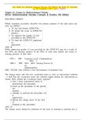
Germ layer contributions – ref to above diagram
- Endoderm – epithelium and associated glands and specialized tissue of gut tube
organs
- Mesoderm (splanchnic) – mesentery, connective tissues, smooth muscle, blood and
lymphatic vessels
- Ectoderm (neural) – enteric nervous system (myenteric and submucosal plexuses)
, 3|Page
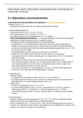
Embryonic folding
Starting week 3 of gastrulation
- Trilaminar plate formed
o Ectoderm
o Endoderm
o Mesoderm
- Layers from top to bottom
o Amniotic cavity
o Ectoderm
o Neural tube/plate
o Notochord
o Intraembryonic mesoderm
o Endoderm
o Yolk sac
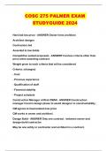
What surrounds the notochord?
- Ventrally – (above the notochord) endoderm
- Dorsally – (below the notochord) ectoderm
- Laterally – (either side of the notochord) lies the mesoderm
- Caudally – (above the notochord end) primitive streak and cloacal membrane
- Rostrally – (below the notochord end) buccopharyngeal membrane and mesoderm region
forming the heart
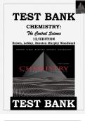
In week 3:
- The mesoderm splits to form paraxial,
intermediate, and lateral plate
mesoderm
o the paraxial mesoderm forms the
dermatome (skin), sclerotome
(bones and ligaments), and
myotome (skeletal muscle)
Embryology of The Gastrointestinal Tract
1) Overview of embryology
2) Embryonic folding
a. Development of the gut tube
b. Development of peritoneum
3) Foregut
a. Components
b. Vascular supply
c. Mesenteries
4) Midgut
a. Components
b. Vascular supply
c. Mesenteries
5) Hindgut
a. Components
b. Vascular supply
c. Mesenteries
6) Intraperitoneal organs
7) Retroperitoneal organs
8) Pathologies
a. Omphalocele
b. Gastroschisis
c. Mackeles diverticulum
,2|Page
Overview of embryology
Germ layer contributions – ref to above diagram
- Endoderm – epithelium and associated glands and specialized tissue of gut tube
organs
- Mesoderm (splanchnic) – mesentery, connective tissues, smooth muscle, blood and
lymphatic vessels
- Ectoderm (neural) – enteric nervous system (myenteric and submucosal plexuses)
, 3|Page
Embryonic folding
Starting week 3 of gastrulation
- Trilaminar plate formed
o Ectoderm
o Endoderm
o Mesoderm
- Layers from top to bottom
o Amniotic cavity
o Ectoderm
o Neural tube/plate
o Notochord
o Intraembryonic mesoderm
o Endoderm
o Yolk sac
What surrounds the notochord?
- Ventrally – (above the notochord) endoderm
- Dorsally – (below the notochord) ectoderm
- Laterally – (either side of the notochord) lies the mesoderm
- Caudally – (above the notochord end) primitive streak and cloacal membrane
- Rostrally – (below the notochord end) buccopharyngeal membrane and mesoderm region
forming the heart
In week 3:
- The mesoderm splits to form paraxial,
intermediate, and lateral plate
mesoderm
o the paraxial mesoderm forms the
dermatome (skin), sclerotome
(bones and ligaments), and
myotome (skeletal muscle)