LECTURE #1
Renal Overview
Did you know that the first successful human organ transplant was a kidney transplant
in 1954? The procedure was between identical twins, so rejection was not a problem.
Kidney transplantation is now the most common organ transplant. Kidneys have a big
reserve capacity. You can lose three-quarters of your kidney function and still be able
to survive. People can also live on one kidney alone and is the reason why people can
be living donors. While the kidneys are an incredible organ, they are not regenerative.
Once part of the kidney has been damaged, it is not repaired by the body. Instead,
having the excess capacity is required, so that when the functional units of the kidneys
are damaged, the nephrons, the kidneys can still maintain function. Interestingly,
approximately 1 in 5000 people are born with only one kidney or are born with only
one functioning kidney. This pathology usually goes undetected, and these individuals
go on to live normal, healthy lives.
*7 func. OVERVIEW OF RENAL FUNCTIONS
1. Regulation of ionic composition of blood
2. Maintenance of blood osmolarity
3. Regulation of blood volume
4. Regulation of blood pressure
5. Regulation of blood pH
6. Excretion of wastes and foreign substances
7. Regulation of blood glucose levels –
gluconeogenesis
8. Production of hormones
KIDNEY ANATOMY
⑭
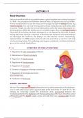
We each have two bean-shaped kidneys that are located posteriorly to the abdomen,
one on each side of your spine, approximately at the level of the 11 th and 12th rib. The
kidneys are technically outside the abdominal cavity, sandwiched between the
membranes that line the abdomen and the bones/muscles of the back, therefore called
-retroperitoneal. The blood supply enters and exits the inner, concave side of each
3
, kidney, as well; the exit of the ureter comes from the inner side of each kidney, which
carries urine from the kidney to the bladder.
THE NEPHRON
The nephron is the functional unit of the kidney, meaning that it is the smallest
structure that can perform the function of an organ, like how the sarcomere is the
functional unit in a muscle. Each kidney has approximately one million nephrons.
2
Plasma and the solutes dissolved within blood plasma filters into nephron and then
this filtered fluid is modified as it travels through the nephron before producing urine.
The nephron is made up of two basic structures, the filtering structure called the renal
corpuscle and the tubule, which processes and modifies the filtered fluid. Capsule
I . -
rencl corpusule (filter)
Nephron ->
glowers
THE RENAL CORPUSCLE- COMPONENTS -> Tubule (process/modifies filterates
The renal corpuscle of every nephron is found in the cortex of the kidney, that is why it
has a granular appearance. The renal corpuscle is made up of two structures, the
capsule and the glomerulus. The nephron capsule is a fluid filled hollow ball-like
structure that surrounds the glomerulus. The capsule is continuous with the beginning
of the first part of the tubule, the proximal convoluted tubule. The cells that make up
the nephron capsule are mostly simple squamous epithelial cells.
G finesterited
The glomerulus is a specialized structure of “leaky” capillaries. The cells that make of
capillaries are called endothelium. The glomerular capillaries have many pores
3
filtering comp (fenestrae) that are so large that basically everything in the blood besides red and white
1) fenestrated cap blood cells can filter out. The endothelium of the glomerular capillaries is fused with
the epithelium of the nephron capsule. The layer of extracellular matrix (ECM) that fuses
2) Basal Lumina these two structures together is called the basal lamina, composed of collagens and
3) podocytes negatively charged glycoproteins. Besides adhering these two structures together, the
basal lamina serves as a rough sieve to further filter and exclude nearly all plasma
↑ proteins from entering the capsule.
A third filtering component of the renal corpuscle are the specialized epithelial cells of
the capsule that contact and surround the glomerular capillaries, called podocytes.
Unlike simple squamous epithelial cells, podocytes have long foot-like processes,
called pedicels, that interlace with each other and around the glomerular capillaries.
Podocytes are also adhered to the basal lamina. These pedicels leave narrow slits
around the capillaries to control the rate of fluid filtration into the capsule. In more
recent years, it has been determined that damage to podocytes can also lead to
protein leak and an increase filtration of fluid, suggesting a role for podocytes in the
exclusion of proteins from entering the capsular space in addition to modifying the rate
of fluid filtration. It is important to note that the epithelial cells that make up the outside
4
Renal Overview
Did you know that the first successful human organ transplant was a kidney transplant
in 1954? The procedure was between identical twins, so rejection was not a problem.
Kidney transplantation is now the most common organ transplant. Kidneys have a big
reserve capacity. You can lose three-quarters of your kidney function and still be able
to survive. People can also live on one kidney alone and is the reason why people can
be living donors. While the kidneys are an incredible organ, they are not regenerative.
Once part of the kidney has been damaged, it is not repaired by the body. Instead,
having the excess capacity is required, so that when the functional units of the kidneys
are damaged, the nephrons, the kidneys can still maintain function. Interestingly,
approximately 1 in 5000 people are born with only one kidney or are born with only
one functioning kidney. This pathology usually goes undetected, and these individuals
go on to live normal, healthy lives.
*7 func. OVERVIEW OF RENAL FUNCTIONS
1. Regulation of ionic composition of blood
2. Maintenance of blood osmolarity
3. Regulation of blood volume
4. Regulation of blood pressure
5. Regulation of blood pH
6. Excretion of wastes and foreign substances
7. Regulation of blood glucose levels –
gluconeogenesis
8. Production of hormones
KIDNEY ANATOMY
⑭
We each have two bean-shaped kidneys that are located posteriorly to the abdomen,
one on each side of your spine, approximately at the level of the 11 th and 12th rib. The
kidneys are technically outside the abdominal cavity, sandwiched between the
membranes that line the abdomen and the bones/muscles of the back, therefore called
-retroperitoneal. The blood supply enters and exits the inner, concave side of each
3
, kidney, as well; the exit of the ureter comes from the inner side of each kidney, which
carries urine from the kidney to the bladder.
THE NEPHRON
The nephron is the functional unit of the kidney, meaning that it is the smallest
structure that can perform the function of an organ, like how the sarcomere is the
functional unit in a muscle. Each kidney has approximately one million nephrons.
2
Plasma and the solutes dissolved within blood plasma filters into nephron and then
this filtered fluid is modified as it travels through the nephron before producing urine.
The nephron is made up of two basic structures, the filtering structure called the renal
corpuscle and the tubule, which processes and modifies the filtered fluid. Capsule
I . -
rencl corpusule (filter)
Nephron ->
glowers
THE RENAL CORPUSCLE- COMPONENTS -> Tubule (process/modifies filterates
The renal corpuscle of every nephron is found in the cortex of the kidney, that is why it
has a granular appearance. The renal corpuscle is made up of two structures, the
capsule and the glomerulus. The nephron capsule is a fluid filled hollow ball-like
structure that surrounds the glomerulus. The capsule is continuous with the beginning
of the first part of the tubule, the proximal convoluted tubule. The cells that make up
the nephron capsule are mostly simple squamous epithelial cells.
G finesterited
The glomerulus is a specialized structure of “leaky” capillaries. The cells that make of
capillaries are called endothelium. The glomerular capillaries have many pores
3
filtering comp (fenestrae) that are so large that basically everything in the blood besides red and white
1) fenestrated cap blood cells can filter out. The endothelium of the glomerular capillaries is fused with
the epithelium of the nephron capsule. The layer of extracellular matrix (ECM) that fuses
2) Basal Lumina these two structures together is called the basal lamina, composed of collagens and
3) podocytes negatively charged glycoproteins. Besides adhering these two structures together, the
basal lamina serves as a rough sieve to further filter and exclude nearly all plasma
↑ proteins from entering the capsule.
A third filtering component of the renal corpuscle are the specialized epithelial cells of
the capsule that contact and surround the glomerular capillaries, called podocytes.
Unlike simple squamous epithelial cells, podocytes have long foot-like processes,
called pedicels, that interlace with each other and around the glomerular capillaries.
Podocytes are also adhered to the basal lamina. These pedicels leave narrow slits
around the capillaries to control the rate of fluid filtration into the capsule. In more
recent years, it has been determined that damage to podocytes can also lead to
protein leak and an increase filtration of fluid, suggesting a role for podocytes in the
exclusion of proteins from entering the capsular space in addition to modifying the rate
of fluid filtration. It is important to note that the epithelial cells that make up the outside
4