DMSU 210 Final Exam Prep
A gallbladder with multiple septate, multiple polyps, and comet-tail artifact on
sonography is consistent with:
Adenomyomatosis
Which of the following is NOT characteristic of gallstones?
posterior enhancement
The sonographic WES sign is characteristic of:
a gallbladder that is completely filled with stones
A 68-year-old woman has right upper quadrant pain. The ultrasound examination
reveals a distended gallbladder with a wall thickness of 1.8 mm. The gallbladder
contains low-level echoes that layer and are gravity dependent and multiple small
echogenic foci that shadow (Figure 2, A and B). The common bile duct measures 10
mm and also contains a small echogenic focus (Figure 2, C) in its distal aspect. What is
the most likely diagnosis?
All of the above; choledocholithiasis, sludge, and cholelithiasis
A 45-year-old man has epigastric pain of 3 week's duration. A right upper quadrant
ultrasound scan reveals multiple small polypoid lesions that arise from the gallbladder
wall. The remaining ultrasound examination results are unremarkable. Subsequent
investigation of the gastrointestinal tract reveals gastroenteritis. What is the most likely
diagnosis for the finding in the gallbladder?
Gastritis
Optimal imaging of the gallbladder generally requires a _____ frequency than that used
to evaluate the right lobe of the liver.
,Higher
A 49-year-old woman is seen at an emergency department with epigastric pain for 1
day, nausea and vomiting, and an elevated temperature. The ultrasound examination
reveals a thick-walled gallbladder and evidence of gallstones (Figure 3, A and B). What
is the most likely diagnosis?
acute cholecystitis
A 36-year-old man is seen with right upper quadrant pain, diarrhea, and fever. History
reveals that the patient recently returned from a 2-week business trip in South America.
An ultrasound examination of the right upper quadrant reveals a 3 cm mass in the right
lobe of the liver that appears to be isoechoic to the liver, with a surrounding hypoechoic
halo. Acoustic enhancement posterior to the mass is also shown (Figure 4, A and B).
What is the most likely diagnosis?
amoebic abscess
Which of the following is NOT characteristic of hepatic abscess?
Hypoechoic mass with thick wall and posterior enhancement
Multiple small masses with echogenic centers
Mass with echogenic foci and dirty shadowing in subhepatic space
NONE of the above
What is the upper limit of normal gallbladder wall thickness if the patient is fasting?
3 mm
Which of the following sonographic characteristics is consistent with pyelonephritis?
Normal-looking kidney
Focal hypoechoic area in renal parenchyma
Renal stones
all of the above
Multiple patient positions are important when scanning the gallbladder because:
, Gallstones are mobile.
Bowel can move out of the way.
The gallbladder moves when the patient moves.
all of the above
Elevation of which of the following laboratory values is most associated with biliary
obstruction?
alkaline phosphatase
A 64-year-old man undergoes an ultrasound scan for right-sided pain to rule out
gallbladder disease. The patient also has a history of autosomal dominant polycystic
kidney disease and bilateral nephrectomy. Ultrasound examination of the right upper
quadrant reveals a normal gallbladder and multiple thin-walled anechoic structures
within the liver (Figure 5). These results are most consistent with what diagnosis?
polycystic disease of the liver
Low-level "sandy" echoes within the gallbladder that move when the patient changes
position but do not exhibit shadowing are consistent with:
gallbladder sludge
The initial test in a patient with cholestasis should be:
liver ultrasound
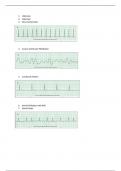
The Doppler waveform in the image above is consistent with:
hepatic vein
The most common cause of hepatocellular disease in the United States is:
alcoholic liver disease
The chronic hallmark of cholestasis is the presence of ascites.
False
A term that means brain disease, damage, or malfunction
A gallbladder with multiple septate, multiple polyps, and comet-tail artifact on
sonography is consistent with:
Adenomyomatosis
Which of the following is NOT characteristic of gallstones?
posterior enhancement
The sonographic WES sign is characteristic of:
a gallbladder that is completely filled with stones
A 68-year-old woman has right upper quadrant pain. The ultrasound examination
reveals a distended gallbladder with a wall thickness of 1.8 mm. The gallbladder
contains low-level echoes that layer and are gravity dependent and multiple small
echogenic foci that shadow (Figure 2, A and B). The common bile duct measures 10
mm and also contains a small echogenic focus (Figure 2, C) in its distal aspect. What is
the most likely diagnosis?
All of the above; choledocholithiasis, sludge, and cholelithiasis
A 45-year-old man has epigastric pain of 3 week's duration. A right upper quadrant
ultrasound scan reveals multiple small polypoid lesions that arise from the gallbladder
wall. The remaining ultrasound examination results are unremarkable. Subsequent
investigation of the gastrointestinal tract reveals gastroenteritis. What is the most likely
diagnosis for the finding in the gallbladder?
Gastritis
Optimal imaging of the gallbladder generally requires a _____ frequency than that used
to evaluate the right lobe of the liver.
,Higher
A 49-year-old woman is seen at an emergency department with epigastric pain for 1
day, nausea and vomiting, and an elevated temperature. The ultrasound examination
reveals a thick-walled gallbladder and evidence of gallstones (Figure 3, A and B). What
is the most likely diagnosis?
acute cholecystitis
A 36-year-old man is seen with right upper quadrant pain, diarrhea, and fever. History
reveals that the patient recently returned from a 2-week business trip in South America.
An ultrasound examination of the right upper quadrant reveals a 3 cm mass in the right
lobe of the liver that appears to be isoechoic to the liver, with a surrounding hypoechoic
halo. Acoustic enhancement posterior to the mass is also shown (Figure 4, A and B).
What is the most likely diagnosis?
amoebic abscess
Which of the following is NOT characteristic of hepatic abscess?
Hypoechoic mass with thick wall and posterior enhancement
Multiple small masses with echogenic centers
Mass with echogenic foci and dirty shadowing in subhepatic space
NONE of the above
What is the upper limit of normal gallbladder wall thickness if the patient is fasting?
3 mm
Which of the following sonographic characteristics is consistent with pyelonephritis?
Normal-looking kidney
Focal hypoechoic area in renal parenchyma
Renal stones
all of the above
Multiple patient positions are important when scanning the gallbladder because:
, Gallstones are mobile.
Bowel can move out of the way.
The gallbladder moves when the patient moves.
all of the above
Elevation of which of the following laboratory values is most associated with biliary
obstruction?
alkaline phosphatase
A 64-year-old man undergoes an ultrasound scan for right-sided pain to rule out
gallbladder disease. The patient also has a history of autosomal dominant polycystic
kidney disease and bilateral nephrectomy. Ultrasound examination of the right upper
quadrant reveals a normal gallbladder and multiple thin-walled anechoic structures
within the liver (Figure 5). These results are most consistent with what diagnosis?
polycystic disease of the liver
Low-level "sandy" echoes within the gallbladder that move when the patient changes
position but do not exhibit shadowing are consistent with:
gallbladder sludge
The initial test in a patient with cholestasis should be:
liver ultrasound
The Doppler waveform in the image above is consistent with:
hepatic vein
The most common cause of hepatocellular disease in the United States is:
alcoholic liver disease
The chronic hallmark of cholestasis is the presence of ascites.
False
A term that means brain disease, damage, or malfunction