Cardiac Dysrhythmias:
Objectives
Normal ECG Components: Correlate with heart’s physiologic events.
ECG Criteria: Identify causes and management of various
dysrhythmias.
Identify Rhythms: Normal sinus rhythm, sinus bradycardia, sinus
tachycardia, premature ventricular contractions, atrial fibrillation,
ventricular tachycardia, ventricular fibrillation, asystole.
Defibrillator Use: Key points.
Defibrillation vs. Cardioversion: Compare
and contrast.
Pacemakers: Use, complications, and nursing
implications.
Continuous Cardiac Monitoring
Electrodes: Place on flat, muscular area;
replace every 24 hours.
Skin Prep: Shave/clip hair, rub skin, cleanse
with alcohol/soap, dry completely.
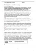
Normal Electrical Conduction
SA Node (Sinus Node): Specialized
muscle cells in the right atrium acting
as the heart’s pacemaker (60-100
beats/min).
AV Node (Atrioventricular Node):
Specialized cardiac fibers at the center
of the heart, slowing and regulating the
signal from the SA node (40-60
beats/min).
Bundle of His: Conduction pathway that transmits impulses from the
AV node to the right and left bundle branches.
Bundle Branches: Right and left branches that carry the conduction
signal.
Purkinje Fibers: Located in the inner ventricular walls, these fibers
conduct impulses, maintaining consistent heart rhythm and
synchronous contractions.
Depolarization: The process where the heart muscle contracts.
Repolarization: The
process where the heart
muscle relaxes.
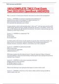
Cardiac Waveforms
PR Interval: 0.12-0.20
seconds.
QT Interval: 0.32-0.40
seconds.
QRS Complex: Less than
0.12 seconds.
P-Wave: Atrial
depolarization (SA node
firing).
, QRS Complex: Ventricular depolarization (AV node relaying signal to
bundle of His and Purkinje fibers).
T-Wave: Ventricular repolarization.
Rhythm Strip Analysis
1. Rate
2. Rhythm regularity
3. Presence of P waves
4. P wave for every QRS?
5. PR interval 0.12-0.20 seconds?
6. QRS complex 0.08-0.12 seconds?
7. QT interval 0.32-0.40 seconds or half R-R distance?
Rhythm Strip Analysis
Amplitude and Voltage: Represented on the vertical axis.
Duration: Represented on the horizontal axis.
Blips: Electrical signals causing deflections on the monitor.
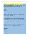
Calculating Heart Rate
6-Second Strip: Each strip has multiple small boxes.
o Large Box: 0.2 seconds.
o Small Box: 0.04 seconds.
Heart Rate Calculation: Count the QRS complexes in a 6-second
strip and multiply by 10.
o Example: 8 QRS complexes in 6 seconds = 8 * 10 = 80 beats per
minute.
Regular vs. Irregular Rhythm
Regular Rhythm: Measure from QRS to QRS. If the distance is
consistent, the rhythm is regular.
Irregular Rhythm: Inconsistent distance between QRS complexes.
P Waves
Presence: Check if P waves are present.
P Wave for Every QRS: Ensure there is one P wave for each QRS
complex.
Regularity: Measure from P wave to P wave to ensure consistency.
PR Interval, QRS Complex, QT Interval
PR Interval: 0.12-0.20 seconds.
QRS Complex: 0.08-0.12 seconds.
Objectives
Normal ECG Components: Correlate with heart’s physiologic events.
ECG Criteria: Identify causes and management of various
dysrhythmias.
Identify Rhythms: Normal sinus rhythm, sinus bradycardia, sinus
tachycardia, premature ventricular contractions, atrial fibrillation,
ventricular tachycardia, ventricular fibrillation, asystole.
Defibrillator Use: Key points.
Defibrillation vs. Cardioversion: Compare
and contrast.
Pacemakers: Use, complications, and nursing
implications.
Continuous Cardiac Monitoring
Electrodes: Place on flat, muscular area;
replace every 24 hours.
Skin Prep: Shave/clip hair, rub skin, cleanse
with alcohol/soap, dry completely.
Normal Electrical Conduction
SA Node (Sinus Node): Specialized
muscle cells in the right atrium acting
as the heart’s pacemaker (60-100
beats/min).
AV Node (Atrioventricular Node):
Specialized cardiac fibers at the center
of the heart, slowing and regulating the
signal from the SA node (40-60
beats/min).
Bundle of His: Conduction pathway that transmits impulses from the
AV node to the right and left bundle branches.
Bundle Branches: Right and left branches that carry the conduction
signal.
Purkinje Fibers: Located in the inner ventricular walls, these fibers
conduct impulses, maintaining consistent heart rhythm and
synchronous contractions.
Depolarization: The process where the heart muscle contracts.
Repolarization: The
process where the heart
muscle relaxes.
Cardiac Waveforms
PR Interval: 0.12-0.20
seconds.
QT Interval: 0.32-0.40
seconds.
QRS Complex: Less than
0.12 seconds.
P-Wave: Atrial
depolarization (SA node
firing).
, QRS Complex: Ventricular depolarization (AV node relaying signal to
bundle of His and Purkinje fibers).
T-Wave: Ventricular repolarization.
Rhythm Strip Analysis
1. Rate
2. Rhythm regularity
3. Presence of P waves
4. P wave for every QRS?
5. PR interval 0.12-0.20 seconds?
6. QRS complex 0.08-0.12 seconds?
7. QT interval 0.32-0.40 seconds or half R-R distance?
Rhythm Strip Analysis
Amplitude and Voltage: Represented on the vertical axis.
Duration: Represented on the horizontal axis.
Blips: Electrical signals causing deflections on the monitor.
Calculating Heart Rate
6-Second Strip: Each strip has multiple small boxes.
o Large Box: 0.2 seconds.
o Small Box: 0.04 seconds.
Heart Rate Calculation: Count the QRS complexes in a 6-second
strip and multiply by 10.
o Example: 8 QRS complexes in 6 seconds = 8 * 10 = 80 beats per
minute.
Regular vs. Irregular Rhythm
Regular Rhythm: Measure from QRS to QRS. If the distance is
consistent, the rhythm is regular.
Irregular Rhythm: Inconsistent distance between QRS complexes.
P Waves
Presence: Check if P waves are present.
P Wave for Every QRS: Ensure there is one P wave for each QRS
complex.
Regularity: Measure from P wave to P wave to ensure consistency.
PR Interval, QRS Complex, QT Interval
PR Interval: 0.12-0.20 seconds.
QRS Complex: 0.08-0.12 seconds.