9-1
Anatomy of the nervous system
The nervous system is broken down into smaller sub-nervous systems to include the central and peripheral
nervous systems.
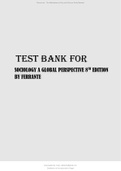
The Central Nervous System is made up of
the brain and spinal cord Peripheral nervous system
The PNS is made up of motor and sensory functions.
❖
Brain The autonomic nervous system : Broken down
into sympathetic and parasympathetic.
❖ The somatic nervous system : Voluntary motor
function control.
❖ Cerebrum: Right and left hemispheres receive sensory info
and relies a motor response.
❖ Cerebellum : Balance
❖ Cerebral cortex: Gray outer layer , 5 lobes, responsible for
conscious activity.
❖ Basal ganglia: Produce smooth voluntary movements. White
mater.
Autonomic nervous system
❖ Diencephalon : Consists of 4 main sections but 2 are most Controls involuntary movements and is broken down into
known for endocrine, sensory, consciousness, and motor sympathetic and parasympathetic systems.
signals to the brain. These two common areas are:
Thalamus: relays sensory impulses. ❖ Controls visceral movement : cardiac, smooth
Hypothalamus: regulates autonomic responses, stress muscle, and glands.
responses, sleep, eating, temperature regulation, fluid balance,
emotions and hormones secreted from the pituitary. ❖ Sympathetic: The fight or Flight response. Blood
❖ Brain Stem: Midbrain, Pons, medulla oblongata gets shunted to vital organs, airway opens up, BP
❖ Cerebellum : coordinates smooth muscle movement and increases, heart rate increases, pupils dilate.
posture. Think Anxiety attack!
❖ Parasympathetic nervous system: Rest and
Spinal cord digest. The body is in a relaxed state. Digestion
and elimination can occur. BP is at a normal level,
heart rate is within normal limits. Think about the
❖ spinal cord ; Provides neuron and synapse networks to post margarita feeling!
produce in voluntary movement to sensory stimulation ( hot
stove, move hand)
Controls body movement and visceral fx, carries sensory information to
the brain and motor fx away from the brain. Extends from the first
cervical to the second lumbar vertebrae, protected by the meninges,
cerebrospinal fluid, and adipose tissue.
❖ Horns : Inner column of grey matter, contains two anterior and
two posteriori or
Posterior horns connect afferently.
Anterior horns move efferently.
❖ Nerve tracts : White matter contains the nerve tracts
Ascending tracts sensory. Descending tract motor ( drive out)
❖ Meninges : The dura mater is a tough and fibrous membrane.
The arachnoid membrane is a delicate membrane and
contains CSF. Pia mater is a vascular membrane.
Subarachnoid space is formed by the arachnoid membrane Somatic nervous system
and the pia mater.
❖ Conducts impulses from the CNS to skeletal
❖ Cerebrospinal fluid : Glowing halo, secreted in the ventricles, muscles. Initiates voluntary movement.
❖
circulates in the subarachnoid space, through the meninges to
❖ consists of afferent nerves or sensory nerves,
the subarachnoid space of the spinal cord where it's and efferent nerves or motor nerves.
absorbed. Acts as a protective cushion aids in the exchange ❖
of nutrients and waste. Normal pressure 50-175 mmhg, ❖ Think sensory going in, like you're walking in a
normal volume 125-150 ml door.
❖
❖ Think motor out, like you're leaving out the
door.
www.SimpleNursing.com
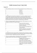
, 9-2 Focused neurological assessm
Assess level of
consciousness Assess orientation
❖ Oriented x3 : understands spoken and written
❖ Full consciousness: Awake and language and responds appropriately.
Alert ❖ Oriented x2: Mild confusion, guesses date, may be
❖ Lethargic: sleeps frequently but able to recognize time of year. My not be able to follow
awakens easily. instructions. May have memory deficits.
❖ Obtunded: extreme drowsiness, ❖ Oriented x1: confused, unable to give date or time,
requires vigorous stimulation to unable to verbalize where or who they are. Has
waken. memory deficits and can be restless or agitated.
❖ Stupor: minimal movement, ❖ Disoriented: patient does not answer appropriately or
responds inappropriately. Is at all. May be hallucinating or agitated. Unable to
awake briefly with vigorous follow directions.
stimulation or painful stimuli.
❖ Comatose: does not respond to
verbal and tactile stimuli. May
respond appropriately to painful
stimuli.
PEERLA
❖ Test pupil response, size, symmetry, shape. They
should be equal and reactive to light.
❖ Shine the penlight into each pupil. Constriction
should be brisk and equal.
❖ Bilateral dilation can be caused by cerebral anoxia or
Assess muscle strength & function anticholinergic medications. Be sure to assess the
client's medication list and other symptoms.
❖ Have the patient move all extremities. ❖ Bilateral constriction can be caused by: intracranial
❖ Have the patient squeeze your fingers.
hemorrhage, opiates, or organophosphates.
❖ Hold your hands up for the patient to push and
pull your hands.
❖ Have the patient hold their arms to their eyes.
Note any drifts. Cranial nerves
❖ Have the patient dorsiflex and plantar flex. ❖ I: olfactory : Smell, have the client identify familiar smells.
❖ Have the patient raise their legs without ❖ II : Optic: Visual acuity, use snellen eye chart, assess
resistance. peripheral vision.
❖ III: Oculomotor: Pupillary reaction, assess PERRLA
Muscle strength scale ❖ IV: Trochlear: Eye movement, patient follows finger without
0 : No muscle movement. movement.
1: Visible muscle movement, no joint movement. ❖ V: Trigeminal: Facial sensation, touch patient's face, have
2: Movement at the Joint but not against gravity. them open their mouth.
3: Movement against gravity but not resistance. ❖ VI: Abducens: Motor function, patient follows finger without
4: Movement against resistance but less than normal moving head.
5: Normal strength. ❖ VII: Facial: Taste and face movement, have patient smile and
puff cheeks, have patient differentiate between sweet and
salty tastes.
Glasgow coma scale ❖ VIII: Acoustic: Hearing and balance, snap fingers close to
patient's ear, have patient stand with feet together, arms at
side and eyes closed for 5 seconds.
❖ IX: Glossopharyngeal: Swallowing and voice, have the
❖ Eye response
patient swallow and say “ah”
Spontaneously 4 ❖ X: Vagus: Gag reflex, use a tongue depressor to swab and
On command 3
elicit a gag reflex.
To pain 2 ❖ XI: Spinal accessory: Neck motion, have patient shrug and
No response 1
turn their head against resistance.
Score____ ❖ XII: Hypoglossal: Tongue movement, have patient stick their
tongue out and move it around.
❖ Verbal response
Alert and oriented 4 ❖ Brain trick to remember the order of cranial nerves.
Confused 3 “ OOO to touch and feel a great velvet super hero”
Inappropriate 2
Incomprehensible 1
score____ Posturing
❖ Decorticate : An abnormal
❖ Motor response
posturing in which a person is stiff
Follows direction 6
with bent arms, clenched fists,
Localizes pain 5
and legs held out straight.
Withdrawal from pain 4
❖ Decerebrate :An abnormal body
Abnormal flexion 3
posture that involves the arms and
Abnormal extension 2
legs being held straight out, the
No response 1
toes being pointed downward,
score____
and the head and neck being
arched backwards
, www.SimpleNursing.com
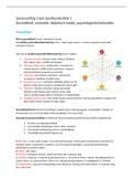
Anatomy of the nervous system
The nervous system is broken down into smaller sub-nervous systems to include the central and peripheral
nervous systems.
The Central Nervous System is made up of
the brain and spinal cord Peripheral nervous system
The PNS is made up of motor and sensory functions.
❖
Brain The autonomic nervous system : Broken down
into sympathetic and parasympathetic.
❖ The somatic nervous system : Voluntary motor
function control.
❖ Cerebrum: Right and left hemispheres receive sensory info
and relies a motor response.
❖ Cerebellum : Balance
❖ Cerebral cortex: Gray outer layer , 5 lobes, responsible for
conscious activity.
❖ Basal ganglia: Produce smooth voluntary movements. White
mater.
Autonomic nervous system
❖ Diencephalon : Consists of 4 main sections but 2 are most Controls involuntary movements and is broken down into
known for endocrine, sensory, consciousness, and motor sympathetic and parasympathetic systems.
signals to the brain. These two common areas are:
Thalamus: relays sensory impulses. ❖ Controls visceral movement : cardiac, smooth
Hypothalamus: regulates autonomic responses, stress muscle, and glands.
responses, sleep, eating, temperature regulation, fluid balance,
emotions and hormones secreted from the pituitary. ❖ Sympathetic: The fight or Flight response. Blood
❖ Brain Stem: Midbrain, Pons, medulla oblongata gets shunted to vital organs, airway opens up, BP
❖ Cerebellum : coordinates smooth muscle movement and increases, heart rate increases, pupils dilate.
posture. Think Anxiety attack!
❖ Parasympathetic nervous system: Rest and
Spinal cord digest. The body is in a relaxed state. Digestion
and elimination can occur. BP is at a normal level,
heart rate is within normal limits. Think about the
❖ spinal cord ; Provides neuron and synapse networks to post margarita feeling!
produce in voluntary movement to sensory stimulation ( hot
stove, move hand)
Controls body movement and visceral fx, carries sensory information to
the brain and motor fx away from the brain. Extends from the first
cervical to the second lumbar vertebrae, protected by the meninges,
cerebrospinal fluid, and adipose tissue.
❖ Horns : Inner column of grey matter, contains two anterior and
two posteriori or
Posterior horns connect afferently.
Anterior horns move efferently.
❖ Nerve tracts : White matter contains the nerve tracts
Ascending tracts sensory. Descending tract motor ( drive out)
❖ Meninges : The dura mater is a tough and fibrous membrane.
The arachnoid membrane is a delicate membrane and
contains CSF. Pia mater is a vascular membrane.
Subarachnoid space is formed by the arachnoid membrane Somatic nervous system
and the pia mater.
❖ Conducts impulses from the CNS to skeletal
❖ Cerebrospinal fluid : Glowing halo, secreted in the ventricles, muscles. Initiates voluntary movement.
❖
circulates in the subarachnoid space, through the meninges to
❖ consists of afferent nerves or sensory nerves,
the subarachnoid space of the spinal cord where it's and efferent nerves or motor nerves.
absorbed. Acts as a protective cushion aids in the exchange ❖
of nutrients and waste. Normal pressure 50-175 mmhg, ❖ Think sensory going in, like you're walking in a
normal volume 125-150 ml door.
❖
❖ Think motor out, like you're leaving out the
door.
www.SimpleNursing.com
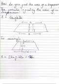
, 9-2 Focused neurological assessm
Assess level of
consciousness Assess orientation
❖ Oriented x3 : understands spoken and written
❖ Full consciousness: Awake and language and responds appropriately.
Alert ❖ Oriented x2: Mild confusion, guesses date, may be
❖ Lethargic: sleeps frequently but able to recognize time of year. My not be able to follow
awakens easily. instructions. May have memory deficits.
❖ Obtunded: extreme drowsiness, ❖ Oriented x1: confused, unable to give date or time,
requires vigorous stimulation to unable to verbalize where or who they are. Has
waken. memory deficits and can be restless or agitated.
❖ Stupor: minimal movement, ❖ Disoriented: patient does not answer appropriately or
responds inappropriately. Is at all. May be hallucinating or agitated. Unable to
awake briefly with vigorous follow directions.
stimulation or painful stimuli.
❖ Comatose: does not respond to
verbal and tactile stimuli. May
respond appropriately to painful
stimuli.
PEERLA
❖ Test pupil response, size, symmetry, shape. They
should be equal and reactive to light.
❖ Shine the penlight into each pupil. Constriction
should be brisk and equal.
❖ Bilateral dilation can be caused by cerebral anoxia or
Assess muscle strength & function anticholinergic medications. Be sure to assess the
client's medication list and other symptoms.
❖ Have the patient move all extremities. ❖ Bilateral constriction can be caused by: intracranial
❖ Have the patient squeeze your fingers.
hemorrhage, opiates, or organophosphates.
❖ Hold your hands up for the patient to push and
pull your hands.
❖ Have the patient hold their arms to their eyes.
Note any drifts. Cranial nerves
❖ Have the patient dorsiflex and plantar flex. ❖ I: olfactory : Smell, have the client identify familiar smells.
❖ Have the patient raise their legs without ❖ II : Optic: Visual acuity, use snellen eye chart, assess
resistance. peripheral vision.
❖ III: Oculomotor: Pupillary reaction, assess PERRLA
Muscle strength scale ❖ IV: Trochlear: Eye movement, patient follows finger without
0 : No muscle movement. movement.
1: Visible muscle movement, no joint movement. ❖ V: Trigeminal: Facial sensation, touch patient's face, have
2: Movement at the Joint but not against gravity. them open their mouth.
3: Movement against gravity but not resistance. ❖ VI: Abducens: Motor function, patient follows finger without
4: Movement against resistance but less than normal moving head.
5: Normal strength. ❖ VII: Facial: Taste and face movement, have patient smile and
puff cheeks, have patient differentiate between sweet and
salty tastes.
Glasgow coma scale ❖ VIII: Acoustic: Hearing and balance, snap fingers close to
patient's ear, have patient stand with feet together, arms at
side and eyes closed for 5 seconds.
❖ IX: Glossopharyngeal: Swallowing and voice, have the
❖ Eye response
patient swallow and say “ah”
Spontaneously 4 ❖ X: Vagus: Gag reflex, use a tongue depressor to swab and
On command 3
elicit a gag reflex.
To pain 2 ❖ XI: Spinal accessory: Neck motion, have patient shrug and
No response 1
turn their head against resistance.
Score____ ❖ XII: Hypoglossal: Tongue movement, have patient stick their
tongue out and move it around.
❖ Verbal response
Alert and oriented 4 ❖ Brain trick to remember the order of cranial nerves.
Confused 3 “ OOO to touch and feel a great velvet super hero”
Inappropriate 2
Incomprehensible 1
score____ Posturing
❖ Decorticate : An abnormal
❖ Motor response
posturing in which a person is stiff
Follows direction 6
with bent arms, clenched fists,
Localizes pain 5
and legs held out straight.
Withdrawal from pain 4
❖ Decerebrate :An abnormal body
Abnormal flexion 3
posture that involves the arms and
Abnormal extension 2
legs being held straight out, the
No response 1
toes being pointed downward,
score____
and the head and neck being
arched backwards
, www.SimpleNursing.com