CARDIOLOGY MCQs 2025 Verified
Cardiology
Exam Guide –
Expert-
Reviewed, Full
Comprehensive
and Up-to-Date
Study Material
for Mastering
Your
Cardiology
Certification
COMPREHENSIVE
EXAM 2025
, CARDIOLOGY MCQs
Q-1
A 62-year-old man is admitted to hospital following a EXPLANATION:
myocardial infarction. Four days after admission he develops Fondaparinux works in a similar way to low-molecular
a further episode of central crushing chest pain. Which is the weight heparin.
best cardiac marker to investigate his chest pain?
ACUTE CORONARY SYNDROME: MANAGEMENT OF NSTEMI
A. LDH NICE produced guidelines in 2013 on the Secondary
B. Troponin I prevention in primary and secondary care for patients
C. Troponin T following a myocardial infarction management of unstable
D. CK-MB angina and non-ST elevation myocardial infarction (NSTEMI).
E. AST These superceded the 2010 guidelines which advocated a risk-
based approach to management which determined whether
ANSWER: drugs such as clopidogrel were given.
CK-MB
All patients should receive
EXPLANATION: • aspirin 300mg
By day four the CK-MB levels should have returned to normal • nitrates or morphine to relieve chest pain if required
from the initial myocardial infarction. If the CK-MB levels are
elevated it would indicate a further coronary event Whilst it is common that non-hypoxic patients receive oxygen
therapy there is little evidence to support this approach. The
CARDIAC ENZYMES AND PROTEIN MARKERS 2008 British Thoracic Society oxygen therapy guidelines advise
Interpretation of the various cardiac enzymes has now largely not giving oxygen unless the patient is hypoxic.
been superceded by the introduction of troponin T and I.
Questions still however commonly appear in exams. Antithrombin treatment. Fondaparinux should be offered to
patients who are not at a high risk of bleeding and who are
Key points for the exam not having angiography within the next 24 hours. If
• myoglobin is the first to rise angiography is likely within 24 hours or a patients creatinine is
• CK-MB is useful to look for reinfarction as it returns to > 265 µmol/l unfractionated heparin should be given.
normal after 2-3 days (troponin T remains elevated for up
to 10 days) Clopidogrel 300mg should be given to all patients and
continued for 12 months.
Begins to rise Peak value Returns to normal
Myoglobin 1-2 hours 6-8 hours 1-2 days Intravenous glycoprotein IIb/IIIa receptor antagonists
CK-MB 2-6 hours 16-20 hours 2-3 days (eptifibatide or tirofiban) should be given to patients who
CK 4-8 hours 16-24 hours 3-4 days have an intermediate or higher risk of adverse cardiovascular
Trop T 4-6 hours 12-24 hours 7-10 days events (predicted 6-month mortality above 3.0%), and who
AST 12-24 hours 36-48 hours 3-4 days are scheduled to undergo angiography within 96 hours of
LDH 24-48 hours 72 hours 8-10 days hospital admission.
Coronary angiography should be considered within 96 hours
Q-2
of first admission to hospital to patients who have a predicted
A patient is admitted with central chest pain and a diagnosis
of non-ST elevation myocardial infarction is made. Aspirin 6-month mortality above 3.0%. It should also be performed as
soon as possible in patients who are clinically unstable.
and fondaparinux are given. What is the mechanism of
action of fondaparinux?
The table below summaries the mechanism of action of drugs
commonly used in the management of acute coronary
A. Reversible direct thrombin inhibitor
syndrome:
B. Glycoprotein IIb/IIIa receptor antagonist
C. Inhibits antithrombin III
D. Inhibits ADP binding to its platelet receptor Medication Mechanism of action
E. Activates antithrombin III Aspirin Antiplatelet - inhibits the production of
thromboxane A2
Clopidogrel Antiplatelet - inhibits ADP binding to its platelet
ANSWER:
receptor
Activates antithrombin III
, Medication Mechanism of action Q-4
Enoxaparin Activates antithrombin III, which in turn Your review a 41-year-old woman. Four months ago she
potentiates the inhibition of coagulation factors developed a deep vein thrombosis and was warfarinised
Xa with a target INR of 2.5. She has presented with a swollen,
Fondaparinux Activates antithrombin III, which in turn tender left calf and a Doppler scan confirms a further deep
potentiates the inhibition of coagulation factors vein thrombosis. Her INR has been above 2.0 for the past
Xa
three months. You organise some investigations to exclude
Bivalirudin Reversible direct thrombin inhibitor
an underlying prothrombotic condition. What should happen
Abciximab, eptifibatide, Glycoprotein IIb/IIIa receptor antagonists
regarding her anticoagulation?
tirofiban
A. Switch to treatment dose low-molecular weight heparin
Q-3
B. Continue on warfarin, continue with INR target of 2.5
An 83-year-old male presents with ischaemic sounding chest
C. Add aspirin 75 mg od
pain that has persisted for the past one hour. A 12-lead ECG
D. Continue on warfarin, increase INR target to 3.0
is performed and shows deep T wave inversion in leads V1
E. Continue on warfarin, increase INR target to 3.5
and V2.
ANSWER:
Which is the most likely implicated coronary artery?
Continue on warfarin, increase INR target to 3.5
A. Left circumflex artery
EXPLANATION:
B. Left main stem artery
WARFARIN
C. Proximal left anterior descending artery
Warfarin is an oral anticoagulant which inhibits the reduction
D. Right coronary artery
of vitamin K to its active hydroquinone form, which in turn
E. Distal left anterior descending artery
acts as a cofactor in the carboxylation of clotting factor II, VII,
IX and X (mnemonic = 1972) and protein C.
ANSWER:
Proximal left anterior descending artery
Indications
• venous thromboembolism: target INR = 2.5, if recurrent
EXPLANATION:
3.5
Wellens' syndrome is an ECG manifestation of critical
• atrial fibrillation, target INR = 2.5
proximal left anterior descending (LAD) coronary artery
• mechanical heart valves, target INR depends on the valve
stenosis in patients with unstable angina. It is characterized
type and location. Mitral valves generally require a higher
by symmetrical, often deep (>2 mm), T wave inversions in the
INR than aortic valves.
anterior precordial leads.
Patients on warfarin are monitored using the INR
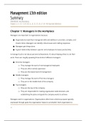
ECG: CORONARY TERRITORIES
(international normalised ration), the ratio of the prothrombin
The table below shows the correlation between ECG changes
time for the patient over the normal prothrombin time.
and coronary territories:
Warfarin has a long half-life and achieving a stable INR may
take several days. There a variety of loading regimes and
ECG changes Coronary artery
computer software is now often used to alter the dose.
Anteroseptal V1-V4 Left anterior descending
Inferior II, III, aVF Right coronary
Factors that may potentiate warfarin
Anterolateral V4-6, I, aVL Left anterior descending or left
• liver disease
circumflex
• P450 enzyme inhibitors, e.g.: amiodarone, ciprofloxacin
Lateral I, aVL +/- V5-6 Left circumflex
• cranberry juice
Posterior Tall R waves V1-2 Usually left circumflex, also right
coronary • drugs which displace warfarin from plasma albumin, e.g.
NSAIDs
• inhibit platelet function: NSAIDs
Side-effects
• haemorrhage
• teratogenic, although can be used in breastfeeding
mothers
• skin necrosis: when warfarin is first started biosynthesis
Diagram showing the correlation between ECG changes and coronary territories in
of protein C is reduced. This results in a temporary
acute coronary syndrome procoagulant state after initially starting warfarin,
, normally avoided by concurrent heparin administration. Causes of fixed split S2
Thrombosis may occur in venules leading to skin necrosis • atrial septal defect
• purple toes
Causes of a widely split S2
Q-5 • deep inspiration
A 71-year-old man with a history of ischaemic heart disease • RBBB
is brought to the Emergency Department following a • pulmonary stenosis
'collapse'. He now feels back to normal. The ECG shows sinus • severe mitral regurgitation
rhythm, 94/min with left bundle branch block. Given the ECG
findings, which one of the following is most likely to be Causes of a reversed (paradoxical) split S2 (P2 occurs before
found on auscultation of the heart? A2)
• LBBB
A. Fixed split S2 • severe aortic stenosis
B. Loud S1 • right ventricular pacing
C. Third heart sound (S3) • WPW type B (causes early P2)
D. Widely split S2 • patent ductus arteriosus
E. Reversed split S2
Q-6
ANSWER: The use of beta-blockers in treating hypertension has
Reversed split S2 declined sharply in the past five years. Which one of the
following best describes the reasons why this has occurred?
EXPLANATION:
Second heart sound (S2)
• Less likely to prevent stroke + potential impairment of
• loud: hypertension glucose tolerance
• soft: AS • Less likely to prevent myocardial infarctions + potential
• fixed split: ASD impairment of glucose tolerance
• reversed split: LBBB • High rate of interactions with other commonly
prescribed medications (e.g. Calcium channel blockers)
HEART SOUNDS: S2 • Increased incidence of reported adverse effects
S2 is caused by the closure of the aortic valve (A2) closely
• Increased incidence of chronic obstructive pulmonary
followed by that of the pulmonary valve (P2)
disease
ANSWER:
Less likely to prevent stroke + potential impairment of glucose
tolerance
EXPLANATION:
This was demonstrated in the Anglo-Scandinavian Cardiac
Outcomes Trial-Blood Pressure Lowering Arm (ASCOT-BPLA).
HYPERTENSION: MANAGEMENT
NICE published updated guidelines for the management of
hypertension in 2011. Some of the key changes include:
• classifying hypertension into stages
• recommending the use of ambulatory blood pressure
monitoring (ABPM) and home blood pressure monitoring
(HBPM)
• calcium channel blockers are now considered superior to
Causes of a loud S2 thiazides
• hypertension: systemic (loud A2) or pulmonary (loud P2) • bendroflumethiazide is no longer the thiazide of choice
• hyperdynamic states
• atrial septal defect without pulmonary hypertension Blood pressure classification
This becomes relevant later in some of the management
Causes of a soft S2 decisions that NICE advocate.
• aortic stenosis
Cardiology
Exam Guide –
Expert-
Reviewed, Full
Comprehensive
and Up-to-Date
Study Material
for Mastering
Your
Cardiology
Certification
COMPREHENSIVE
EXAM 2025
, CARDIOLOGY MCQs
Q-1
A 62-year-old man is admitted to hospital following a EXPLANATION:
myocardial infarction. Four days after admission he develops Fondaparinux works in a similar way to low-molecular
a further episode of central crushing chest pain. Which is the weight heparin.
best cardiac marker to investigate his chest pain?
ACUTE CORONARY SYNDROME: MANAGEMENT OF NSTEMI
A. LDH NICE produced guidelines in 2013 on the Secondary
B. Troponin I prevention in primary and secondary care for patients
C. Troponin T following a myocardial infarction management of unstable
D. CK-MB angina and non-ST elevation myocardial infarction (NSTEMI).
E. AST These superceded the 2010 guidelines which advocated a risk-
based approach to management which determined whether
ANSWER: drugs such as clopidogrel were given.
CK-MB
All patients should receive
EXPLANATION: • aspirin 300mg
By day four the CK-MB levels should have returned to normal • nitrates or morphine to relieve chest pain if required
from the initial myocardial infarction. If the CK-MB levels are
elevated it would indicate a further coronary event Whilst it is common that non-hypoxic patients receive oxygen
therapy there is little evidence to support this approach. The
CARDIAC ENZYMES AND PROTEIN MARKERS 2008 British Thoracic Society oxygen therapy guidelines advise
Interpretation of the various cardiac enzymes has now largely not giving oxygen unless the patient is hypoxic.
been superceded by the introduction of troponin T and I.
Questions still however commonly appear in exams. Antithrombin treatment. Fondaparinux should be offered to
patients who are not at a high risk of bleeding and who are
Key points for the exam not having angiography within the next 24 hours. If
• myoglobin is the first to rise angiography is likely within 24 hours or a patients creatinine is
• CK-MB is useful to look for reinfarction as it returns to > 265 µmol/l unfractionated heparin should be given.
normal after 2-3 days (troponin T remains elevated for up
to 10 days) Clopidogrel 300mg should be given to all patients and
continued for 12 months.
Begins to rise Peak value Returns to normal
Myoglobin 1-2 hours 6-8 hours 1-2 days Intravenous glycoprotein IIb/IIIa receptor antagonists
CK-MB 2-6 hours 16-20 hours 2-3 days (eptifibatide or tirofiban) should be given to patients who
CK 4-8 hours 16-24 hours 3-4 days have an intermediate or higher risk of adverse cardiovascular
Trop T 4-6 hours 12-24 hours 7-10 days events (predicted 6-month mortality above 3.0%), and who
AST 12-24 hours 36-48 hours 3-4 days are scheduled to undergo angiography within 96 hours of
LDH 24-48 hours 72 hours 8-10 days hospital admission.
Coronary angiography should be considered within 96 hours
Q-2
of first admission to hospital to patients who have a predicted
A patient is admitted with central chest pain and a diagnosis
of non-ST elevation myocardial infarction is made. Aspirin 6-month mortality above 3.0%. It should also be performed as
soon as possible in patients who are clinically unstable.
and fondaparinux are given. What is the mechanism of
action of fondaparinux?
The table below summaries the mechanism of action of drugs
commonly used in the management of acute coronary
A. Reversible direct thrombin inhibitor
syndrome:
B. Glycoprotein IIb/IIIa receptor antagonist
C. Inhibits antithrombin III
D. Inhibits ADP binding to its platelet receptor Medication Mechanism of action
E. Activates antithrombin III Aspirin Antiplatelet - inhibits the production of
thromboxane A2
Clopidogrel Antiplatelet - inhibits ADP binding to its platelet
ANSWER:
receptor
Activates antithrombin III
, Medication Mechanism of action Q-4
Enoxaparin Activates antithrombin III, which in turn Your review a 41-year-old woman. Four months ago she
potentiates the inhibition of coagulation factors developed a deep vein thrombosis and was warfarinised
Xa with a target INR of 2.5. She has presented with a swollen,
Fondaparinux Activates antithrombin III, which in turn tender left calf and a Doppler scan confirms a further deep
potentiates the inhibition of coagulation factors vein thrombosis. Her INR has been above 2.0 for the past
Xa
three months. You organise some investigations to exclude
Bivalirudin Reversible direct thrombin inhibitor
an underlying prothrombotic condition. What should happen
Abciximab, eptifibatide, Glycoprotein IIb/IIIa receptor antagonists
regarding her anticoagulation?
tirofiban
A. Switch to treatment dose low-molecular weight heparin
Q-3
B. Continue on warfarin, continue with INR target of 2.5
An 83-year-old male presents with ischaemic sounding chest
C. Add aspirin 75 mg od
pain that has persisted for the past one hour. A 12-lead ECG
D. Continue on warfarin, increase INR target to 3.0
is performed and shows deep T wave inversion in leads V1
E. Continue on warfarin, increase INR target to 3.5
and V2.
ANSWER:
Which is the most likely implicated coronary artery?
Continue on warfarin, increase INR target to 3.5
A. Left circumflex artery
EXPLANATION:
B. Left main stem artery
WARFARIN
C. Proximal left anterior descending artery
Warfarin is an oral anticoagulant which inhibits the reduction
D. Right coronary artery
of vitamin K to its active hydroquinone form, which in turn
E. Distal left anterior descending artery
acts as a cofactor in the carboxylation of clotting factor II, VII,
IX and X (mnemonic = 1972) and protein C.
ANSWER:
Proximal left anterior descending artery
Indications
• venous thromboembolism: target INR = 2.5, if recurrent
EXPLANATION:
3.5
Wellens' syndrome is an ECG manifestation of critical
• atrial fibrillation, target INR = 2.5
proximal left anterior descending (LAD) coronary artery
• mechanical heart valves, target INR depends on the valve
stenosis in patients with unstable angina. It is characterized
type and location. Mitral valves generally require a higher
by symmetrical, often deep (>2 mm), T wave inversions in the
INR than aortic valves.
anterior precordial leads.
Patients on warfarin are monitored using the INR
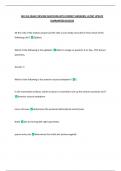
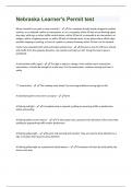
ECG: CORONARY TERRITORIES
(international normalised ration), the ratio of the prothrombin
The table below shows the correlation between ECG changes
time for the patient over the normal prothrombin time.
and coronary territories:
Warfarin has a long half-life and achieving a stable INR may
take several days. There a variety of loading regimes and
ECG changes Coronary artery
computer software is now often used to alter the dose.
Anteroseptal V1-V4 Left anterior descending
Inferior II, III, aVF Right coronary
Factors that may potentiate warfarin
Anterolateral V4-6, I, aVL Left anterior descending or left
• liver disease
circumflex
• P450 enzyme inhibitors, e.g.: amiodarone, ciprofloxacin
Lateral I, aVL +/- V5-6 Left circumflex
• cranberry juice
Posterior Tall R waves V1-2 Usually left circumflex, also right
coronary • drugs which displace warfarin from plasma albumin, e.g.
NSAIDs
• inhibit platelet function: NSAIDs
Side-effects
• haemorrhage
• teratogenic, although can be used in breastfeeding
mothers
• skin necrosis: when warfarin is first started biosynthesis
Diagram showing the correlation between ECG changes and coronary territories in
of protein C is reduced. This results in a temporary
acute coronary syndrome procoagulant state after initially starting warfarin,
, normally avoided by concurrent heparin administration. Causes of fixed split S2
Thrombosis may occur in venules leading to skin necrosis • atrial septal defect
• purple toes
Causes of a widely split S2
Q-5 • deep inspiration
A 71-year-old man with a history of ischaemic heart disease • RBBB
is brought to the Emergency Department following a • pulmonary stenosis
'collapse'. He now feels back to normal. The ECG shows sinus • severe mitral regurgitation
rhythm, 94/min with left bundle branch block. Given the ECG
findings, which one of the following is most likely to be Causes of a reversed (paradoxical) split S2 (P2 occurs before
found on auscultation of the heart? A2)
• LBBB
A. Fixed split S2 • severe aortic stenosis
B. Loud S1 • right ventricular pacing
C. Third heart sound (S3) • WPW type B (causes early P2)
D. Widely split S2 • patent ductus arteriosus
E. Reversed split S2
Q-6
ANSWER: The use of beta-blockers in treating hypertension has
Reversed split S2 declined sharply in the past five years. Which one of the
following best describes the reasons why this has occurred?
EXPLANATION:
Second heart sound (S2)
• Less likely to prevent stroke + potential impairment of
• loud: hypertension glucose tolerance
• soft: AS • Less likely to prevent myocardial infarctions + potential
• fixed split: ASD impairment of glucose tolerance
• reversed split: LBBB • High rate of interactions with other commonly
prescribed medications (e.g. Calcium channel blockers)
HEART SOUNDS: S2 • Increased incidence of reported adverse effects
S2 is caused by the closure of the aortic valve (A2) closely
• Increased incidence of chronic obstructive pulmonary
followed by that of the pulmonary valve (P2)
disease
ANSWER:
Less likely to prevent stroke + potential impairment of glucose
tolerance
EXPLANATION:
This was demonstrated in the Anglo-Scandinavian Cardiac
Outcomes Trial-Blood Pressure Lowering Arm (ASCOT-BPLA).
HYPERTENSION: MANAGEMENT
NICE published updated guidelines for the management of
hypertension in 2011. Some of the key changes include:
• classifying hypertension into stages
• recommending the use of ambulatory blood pressure
monitoring (ABPM) and home blood pressure monitoring
(HBPM)
• calcium channel blockers are now considered superior to
Causes of a loud S2 thiazides
• hypertension: systemic (loud A2) or pulmonary (loud P2) • bendroflumethiazide is no longer the thiazide of choice
• hyperdynamic states
• atrial septal defect without pulmonary hypertension Blood pressure classification
This becomes relevant later in some of the management
Causes of a soft S2 decisions that NICE advocate.
• aortic stenosis