Study Guide for Quiz 6
• Identify the waveforms in a normal electrocardiogram (ECG), and relate the ECG
waveforms to atrial and ventricular depolarization and repolarization and to the
activity of the conduction system.
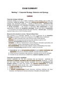
- Small, initial P wave represents depolarization of all cells within atria except SA node; Flat
line before P wave represents depolarization of SA node.
- Large QRS complex represents ventricular depolarization; actually three separate waves;
Q wave is first downward deflection; R is large upward deflection; S is following downward
deflection
- Small T wave occurs after S wave of QRS complex; represents ventricular repolarization; T
wave is an upward deflection under normal conditions
• Describe the phases of the cardiac cycle.
- Systole – Contraction
- Diastole – Contraction
- Ventricular filling
- a. LEFT ATRIA BLOOD PRESSURE is lower than the BLOOD PRESSURE of the pulmonary
trunk, aorta. so blood enters the left atrium.
- b. LA BP is greater than LV BP. Thus the mitral valve is open and blood enters the LV. c. LV
BP is less than aortic BP. As a result, blood tries to back flow from the aorta into the LV and
this forces the aortic semilunar valve closed.
12
, - d. Neither atrial nor ventricular muscle is contracting. Both are in diastole.
- e. About 80% of the ultimate ventricular volume will enter in this passive manner.
- f. Near the end of filling, while the LV is still relaxing, the LA depolarizes and contracts.
This pushes roughly the final 20% of blood into the LV.
- g. LV now has the max volume it will contain during this cycle.- Volume of blood in the
ventricles at the end of diastole. This is the end diastolic volume (EDV). (= 120mL). For the
rest of the cycle, the LA will be in diastole.
- Isovolumetric contraction
- a. LV depolarizes, contracts, and LV BP rises and almost immediately exceeds LA BP.
- b. Blood is pushed upward shutting the mitral valve– creating the 1st heart sound (LUB).
- c. However, the opening of the aortic semilunar valve requires much more pressure than
was necessary to close the mitral valve.
- d. So after the mitral valve is shut, the LV continues to contract and its BP rises, but until
LV BP exceeds aortic BP the aortic semilunar valve remains shut.
- e. Thus, during this period the AV and semilunar valves are shut and the volume within the
LV is not changing. this phase is known as “iso” “volumetric” contraction.
- Ventricular ejection
- a. LV BP now exceeds aortic BP (80mmHg), the semilunar valve is forced open, and blood is
ejected from the LV into the ascending aorta.
- b. Not all of the blood in the LV is ejected. The amount remaining after ventricular
contraction is known as the end systolic volume (ESV). A typical value is 70mL. This gives a
reserve that could also be ejected if needed (e.g., during exercise).
- c. A more vigorous contraction will decrease ESV and increase SV.
- Isovolumetric relaxation
- a. LV stops contracting, its BP falls and quickly becomes less than aortic BP and blood tries
to back flow, which shuts the semilunar valve– creating the 2nd heart sound (DUB).
- b. It takes more time for LV BP to drop below LA BP – and cause the mitral valve to open.
- c. During this time, as LV BP is falling, the AV and semilunar valves are shut and LV volume
is not changing.
- d. Once LV BP falls below LA BP (which is rising as blood returns to the heart), the mitral
valve will open and the cycle will begin anew with another round of ventricular filling.
- E. Blood is not entering nor ejected from ventricles – leaving their volume remaining
constant. – doesn’t change.
• Relate the opening and closing of specific heart valves in each phase of the cardiac
cycle to pressure changes in the heart chambers.
State of AV Valves State of Semilunar Valves
Isovolumetric Contraction closed closed
Isovolumetric Relaxation closed closed
Ventricular Ejection closed open
12
• Identify the waveforms in a normal electrocardiogram (ECG), and relate the ECG
waveforms to atrial and ventricular depolarization and repolarization and to the
activity of the conduction system.
- Small, initial P wave represents depolarization of all cells within atria except SA node; Flat
line before P wave represents depolarization of SA node.
- Large QRS complex represents ventricular depolarization; actually three separate waves;
Q wave is first downward deflection; R is large upward deflection; S is following downward
deflection
- Small T wave occurs after S wave of QRS complex; represents ventricular repolarization; T
wave is an upward deflection under normal conditions
• Describe the phases of the cardiac cycle.
- Systole – Contraction
- Diastole – Contraction
- Ventricular filling
- a. LEFT ATRIA BLOOD PRESSURE is lower than the BLOOD PRESSURE of the pulmonary
trunk, aorta. so blood enters the left atrium.
- b. LA BP is greater than LV BP. Thus the mitral valve is open and blood enters the LV. c. LV
BP is less than aortic BP. As a result, blood tries to back flow from the aorta into the LV and
this forces the aortic semilunar valve closed.
12
, - d. Neither atrial nor ventricular muscle is contracting. Both are in diastole.
- e. About 80% of the ultimate ventricular volume will enter in this passive manner.
- f. Near the end of filling, while the LV is still relaxing, the LA depolarizes and contracts.
This pushes roughly the final 20% of blood into the LV.
- g. LV now has the max volume it will contain during this cycle.- Volume of blood in the
ventricles at the end of diastole. This is the end diastolic volume (EDV). (= 120mL). For the
rest of the cycle, the LA will be in diastole.
- Isovolumetric contraction
- a. LV depolarizes, contracts, and LV BP rises and almost immediately exceeds LA BP.
- b. Blood is pushed upward shutting the mitral valve– creating the 1st heart sound (LUB).
- c. However, the opening of the aortic semilunar valve requires much more pressure than
was necessary to close the mitral valve.
- d. So after the mitral valve is shut, the LV continues to contract and its BP rises, but until
LV BP exceeds aortic BP the aortic semilunar valve remains shut.
- e. Thus, during this period the AV and semilunar valves are shut and the volume within the
LV is not changing. this phase is known as “iso” “volumetric” contraction.
- Ventricular ejection
- a. LV BP now exceeds aortic BP (80mmHg), the semilunar valve is forced open, and blood is
ejected from the LV into the ascending aorta.
- b. Not all of the blood in the LV is ejected. The amount remaining after ventricular
contraction is known as the end systolic volume (ESV). A typical value is 70mL. This gives a
reserve that could also be ejected if needed (e.g., during exercise).
- c. A more vigorous contraction will decrease ESV and increase SV.
- Isovolumetric relaxation
- a. LV stops contracting, its BP falls and quickly becomes less than aortic BP and blood tries
to back flow, which shuts the semilunar valve– creating the 2nd heart sound (DUB).
- b. It takes more time for LV BP to drop below LA BP – and cause the mitral valve to open.
- c. During this time, as LV BP is falling, the AV and semilunar valves are shut and LV volume
is not changing.
- d. Once LV BP falls below LA BP (which is rising as blood returns to the heart), the mitral
valve will open and the cycle will begin anew with another round of ventricular filling.
- E. Blood is not entering nor ejected from ventricles – leaving their volume remaining
constant. – doesn’t change.
• Relate the opening and closing of specific heart valves in each phase of the cardiac
cycle to pressure changes in the heart chambers.
State of AV Valves State of Semilunar Valves
Isovolumetric Contraction closed closed
Isovolumetric Relaxation closed closed
Ventricular Ejection closed open
12