Lecture 10: 11/12
Applica'ons
Protein iden*fica*on
- Past: Purifica-on of protein -> digest -> Edman degrada-on: laborious!
- Top-down strategy (1):
o Determine MW of the protein (if resolu-on is high enough) -> sufficient when no
PTM present.
- BoHom-up strategy (2):
o Trypsinise protein and iden-fy on the basis of PMF and/or PFF (or de novo)
o ANer digest only a limited number of pep-des has PTM, hence iden-fica-on is
possible.
o Top down
- Protein not know -> de novo sequencing or sequencing parts of the protein and then
produce oligonucleo-des for further cloning
- Iden-fica-on without prior separa-on can be performed on less complex protein
mixtures (max. 5-6 proteins/sample).
o Mixture not too complex -> immediately introduce your sample in the mass spect
without prior isola-on or purifica-on and immediately perform iden-fica-on on
the complex
- Sample is more complex -> purifica-on by chromatography or gel electrophoresis
Detec*on and characteriza*on of muta*ons
- Muta-ons result in MW differences, varying between 0.0364 Da (Gln/lys) and 129.0578
(Gly/Trp)
- Three steps:
o (Protein MW determina-on (high resolu-on and accuracy necessary))
o Enzyma-c digest for determina-on of mutated pep-de (based upon MW
difference)
o Sequence determina-on of mutated pep-de
- One AA replaced by another AA -> MW of pep-de will differ -> you can look for
pep-des that differ according to the molecular weight of that pep-de in databases
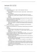
- Example Separa-ng alfa and beta chains
o MALDI-TOF: detects muta-ons in the b-chain (mutant chain is 14 Da heavier as
compared to na-ve chain)
o ANer tryp-c digest of the β chains MALDI-TOF shows two new pep-des: T9m and
T8+T9m (results from missed cleavage).
m/z T9m = 1683.90 = 14.01 Da heavier as compared to normal pep-de T9 =
1669.89.
o Since the difference of 14.01 Da corresponds to 6 different muta-ons (G/A, S/T,
V/I, V/L, N/Q of D/E; see table) and since al these AAs (G, S, V, N en D) are present
in the pep-de T9 (AZ sequence 67-82), the nature of the muta-on cannot solely
be determined from the mass difference.
o MS/MS is necessary for sequence determina-on of T9m. Conclusion: Asp79
replaced by Glu
, o Some pep-des are ok and some of them are split up -> normal and mutant
present.
o Muta-on in B chain -> it looks like you have a slit of the B globin chain
o Pep-des; some are ok, and other are spliHed
§ T9; normal and mutant form are present
§ T8 + T9: trypsiniza-on didn’t occur
o Difference of 14.01 Da -> many of the mutant form are present in the pep-de T9
-> we s-ll don’t know which AA have been replaced -> sequence the T9 mutant
pep-de -> Asp79 replaced by Glu
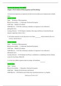
Verifica*on of structure and purity of proteins and pep*des
- Series of peaks -> protein of 14.590 Da -> p18 (MW = 14.589 Da)
- However: two other series are present:
o Small T one (8%): MW = 12.651 Da. Corresponds to C-terminal part of p18 aNer
splicing at posi-on 111-11
o Small D one (3%): MW = 29.175 Da. Corresponds to dimer generated aNer
disulfide bridge forma-on between two p18 proteins.
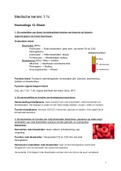
Non-covalent protein complexes and 3D structure informa*on
Na#ve-denatured:
- Distribu-on of charges in denatured protein is broader + more charges are present.
This is the result of the exposi-on of larger protein surfaces allowing the ioniza-on of
plenty of basic AAs.
- Example: acidic denatura-on of human myoglobin in func-on of -me -> 3 species are
observed:
o na-ve hMb (haem + myoglobin)
o denatured hMb (haem + denatured myoglobin)
o denatured aMb (denatured apomyoglobin = without haem moiety)
- Simple method to find out something about the structure of the protein. When the
proteins is denatured -> protein is unfolded and many more AA are acceptable for
protona-on -> will have more charges as compared to the unfolded proteins
- You know something about the folding of the protein compared to the complete folded
form.
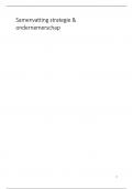
Accessibility
- Accessibility of a certain AA for the solvent can be inves-gated by reac-on of the
protein with reagents that cause irreversible reac-ons, specific for the AA side chain.
Number and posi-on of the modified AAs determines posi-on in the protein 3D
structure.
o When the has AA has covalently bonded groups the MW will increase -> will be at
the surface of the proteins
Internal sites
- Internal sites are protected by bulky groups and will be trypsinized less easy. Hence the
masses of the tryp-c pep-des will give informa-on about the loca-on of some K and
R residues.
o Treat sample, your purified protein with limited amount trypsin -> will cut at the
surface of the protein but there is not enough trypsine to further cut the protein,
only the outside of the protein will be trypsinized -> which pep-de will appear?
These lysins and arginines will be at the outside of the protein
Applica'ons
Protein iden*fica*on
- Past: Purifica-on of protein -> digest -> Edman degrada-on: laborious!
- Top-down strategy (1):
o Determine MW of the protein (if resolu-on is high enough) -> sufficient when no
PTM present.
- BoHom-up strategy (2):
o Trypsinise protein and iden-fy on the basis of PMF and/or PFF (or de novo)
o ANer digest only a limited number of pep-des has PTM, hence iden-fica-on is
possible.
o Top down
- Protein not know -> de novo sequencing or sequencing parts of the protein and then
produce oligonucleo-des for further cloning
- Iden-fica-on without prior separa-on can be performed on less complex protein
mixtures (max. 5-6 proteins/sample).
o Mixture not too complex -> immediately introduce your sample in the mass spect
without prior isola-on or purifica-on and immediately perform iden-fica-on on
the complex
- Sample is more complex -> purifica-on by chromatography or gel electrophoresis
Detec*on and characteriza*on of muta*ons
- Muta-ons result in MW differences, varying between 0.0364 Da (Gln/lys) and 129.0578
(Gly/Trp)
- Three steps:
o (Protein MW determina-on (high resolu-on and accuracy necessary))
o Enzyma-c digest for determina-on of mutated pep-de (based upon MW
difference)
o Sequence determina-on of mutated pep-de
- One AA replaced by another AA -> MW of pep-de will differ -> you can look for
pep-des that differ according to the molecular weight of that pep-de in databases
- Example Separa-ng alfa and beta chains
o MALDI-TOF: detects muta-ons in the b-chain (mutant chain is 14 Da heavier as
compared to na-ve chain)
o ANer tryp-c digest of the β chains MALDI-TOF shows two new pep-des: T9m and
T8+T9m (results from missed cleavage).
m/z T9m = 1683.90 = 14.01 Da heavier as compared to normal pep-de T9 =
1669.89.
o Since the difference of 14.01 Da corresponds to 6 different muta-ons (G/A, S/T,
V/I, V/L, N/Q of D/E; see table) and since al these AAs (G, S, V, N en D) are present
in the pep-de T9 (AZ sequence 67-82), the nature of the muta-on cannot solely
be determined from the mass difference.
o MS/MS is necessary for sequence determina-on of T9m. Conclusion: Asp79
replaced by Glu
, o Some pep-des are ok and some of them are split up -> normal and mutant
present.
o Muta-on in B chain -> it looks like you have a slit of the B globin chain
o Pep-des; some are ok, and other are spliHed
§ T9; normal and mutant form are present
§ T8 + T9: trypsiniza-on didn’t occur
o Difference of 14.01 Da -> many of the mutant form are present in the pep-de T9
-> we s-ll don’t know which AA have been replaced -> sequence the T9 mutant
pep-de -> Asp79 replaced by Glu
Verifica*on of structure and purity of proteins and pep*des
- Series of peaks -> protein of 14.590 Da -> p18 (MW = 14.589 Da)
- However: two other series are present:
o Small T one (8%): MW = 12.651 Da. Corresponds to C-terminal part of p18 aNer
splicing at posi-on 111-11
o Small D one (3%): MW = 29.175 Da. Corresponds to dimer generated aNer
disulfide bridge forma-on between two p18 proteins.
Non-covalent protein complexes and 3D structure informa*on
Na#ve-denatured:
- Distribu-on of charges in denatured protein is broader + more charges are present.
This is the result of the exposi-on of larger protein surfaces allowing the ioniza-on of
plenty of basic AAs.
- Example: acidic denatura-on of human myoglobin in func-on of -me -> 3 species are
observed:
o na-ve hMb (haem + myoglobin)
o denatured hMb (haem + denatured myoglobin)
o denatured aMb (denatured apomyoglobin = without haem moiety)
- Simple method to find out something about the structure of the protein. When the
proteins is denatured -> protein is unfolded and many more AA are acceptable for
protona-on -> will have more charges as compared to the unfolded proteins
- You know something about the folding of the protein compared to the complete folded
form.
Accessibility
- Accessibility of a certain AA for the solvent can be inves-gated by reac-on of the
protein with reagents that cause irreversible reac-ons, specific for the AA side chain.
Number and posi-on of the modified AAs determines posi-on in the protein 3D
structure.
o When the has AA has covalently bonded groups the MW will increase -> will be at
the surface of the proteins
Internal sites
- Internal sites are protected by bulky groups and will be trypsinized less easy. Hence the
masses of the tryp-c pep-des will give informa-on about the loca-on of some K and
R residues.
o Treat sample, your purified protein with limited amount trypsin -> will cut at the
surface of the protein but there is not enough trypsine to further cut the protein,
only the outside of the protein will be trypsinized -> which pep-de will appear?
These lysins and arginines will be at the outside of the protein