9-1
Various secondary lymphoid organs are organised with distinct areas for B
and T cells B cell and T cell zones. The SLOs also contain macrophages,
DCs and nonleukocyte stromal cells.
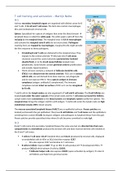
Spleen: Specialized for capture of antigens that enter the blood stream
lymphoid tissue is called the white pulp. The white pulp is split off from the
red pulp by the marginal sinus. The marginal sinus is rich in macrophages
and contains the marginal zone B cells (do not recirculate). Pathogens
reaching here are trapped by macrophages, marginal B cells might provide
the first response to these pathogens.
Circulating B and T cells are delivered to the marginal sinus. They
migrate to the central arteriole bifurcate into T cell zones
clustered around the central arteriole (periarteriolar lymhoid
sheath/PALS) or to the B cell zones/follicles located more
peripherally. Some folicles contain germinal centres (proliferation
and somatic hypermutation).
The B cell zone contains a network of follicular dendritic cells
(FDCs) most distant from the central arteriole. FDCs are in contact
with B cells, are not derived from bone marrow, not phagocytic
and do not express MHC II. They capture antigen in immune
complexes (antigen, antibody & complement). The immune
complexes remain intact on surface of FDC so it can be recognised
by B cells.
T and B cells in the lymph nodes are also organised in T cell and B cell zones. The B cell follicles are
located just under the outer capsule of the lymph node and the T cell zones surround the follicles.
Lymph nodes have connections to the blood system and lymphatic system (unlike the spleen). The
marginal sinus brings the antigen and DCs with antigen. T and B cells enter the lymph node via high
endothelial venules (HEV, blood vessels).
The mucosa associated lymphoid tissues (MALT) are on epithelial surfaces. Peyers patches are
located just beneath the gut epithelium They have B cell follicles and T cell zones and the epithelium
overlying them contain M cells (transport antigens and pathogens to lymphoid tissue from the gut).
Peyers patches provide specialized sites where B cells become commited to make IgA.
9-3
B and T cells come into secondary lymphoid tissues the same way but are directed into their own
compartments by chemokines produced by stromal cells and bone marrow derived cells resident in
the B and T cell zones.
T cells to T cell zone: CCL19 (resident DCs) and CCL21 (produced by stromal cells, displayed
on endothelial cells in HEVs or DCs) bind the receptor CCR7.
o DCs also express CCR7 and localise to T cell zones.
B cells to follicle: Express CCR7 go to HEV. B cells produce LT development FDCs
produce CXCL13 which attracts B cells by CXCR5.
o T follicular helper cells also express CXCR5 (upon activation by antigen) enter B
cell follicles and help form germinal center.
, 9-4
Naive T cells in blood lymph nodes/spleen/mucosa associated lymphoid tissues blood. Allows
them to contact many DCs & sample peptide:MHC complexes. Within hours of arrival, naive T cells
that did not encounter their antigen leave the lymphoid tissue and re-enter the bloodstream.
When a naive T cell encounters its antigen stays in T cell zone proliferation, clonal expansion,
differentiation effector/memory T cell. At the end of this, they exit the lymphoid tissue and re
enter the blood stream migrate to site of infection. Some effector T cells go to B cell zones &
participate in germinal center response (Tfh).
Naive T cell screening for antigen very efficient. Within 48 hours all cells checked.
9-5/6
Migration of naive T cells to secondary lymphoid tissues
depends on binding to HEVs through cell-cell interactions due
to cell adhesion molecules main class are selectins, integrins,
members of immunoglobulin superfamily and mucin-like
molecules.
Rolling: L-selectins on naive T cells initiates a light
attachment to the wall of the HEV so the T cell can roll
along the endothelial surface. CD34 and GlyCAM1 in
lymph nodes & MAdCAM-1 in mucosae.
o P-selectin and E-selectin are expressed on vascular endothelium at sites of infection
and serve to recruit effector cells to infected tissue.
o Selectins are cell surface molecules with a common core structure but have different
lectin like domains on their extracellular portion. The selectin binds to a cell surface
carbohydrate.
Activation: Signalling by chemokines (CCL21) activates integrins on leukocytes to bind tightly
to the vascular wall, LFA-1 becomes activated
Adhesion: Conformation of integrins (LFA-1) are changed by chemokines leukocytes and
integrins bind tightly. LFA is activated increased affinity for ICAM1/2.
Diapedesis: Chemokines bind to
proteoglycans in extracellular matrix
HEVs gradient recognised by
receptors on T cells. CCL21 from HEV
cells, stromal cells and DCs in
lymphoid tissues bind to CCR7 on
T cell activation intracellular
receptor associated G protein
subunit Galfai stronger binding to
integrin.
Once naive T cells arrived in the T cell zone CCR7 directs retention. Attracted to DCs that produce
CCL21 and CCL19 in T cell zone. They scan DCs for peptide:MHC complexes if they bind trapped, if
not, they leave.
Integrin consists of large alfa chain that pairs noncovalently with a smaller beta chain. All T cells
express integrin alfaLbeta2 LFA-1. LFA-1 enables migration of naive and effector T cells out of the
blood (also present on macrophages and neutrophils). It is also important in the adhesion of naive

