Lecture 7 Abbreviation Key:
Monday, August 19, 2019 10:00 AM b/c = because
b/w = between
w/ = with
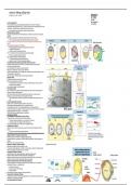
• Interaction w/ VegT and B-catenin. expt = experiment
○ B-catenin left side. ex. = example
○ VegT bottom side.
• VegT induces LOW nodal -> ventral mesoderm
• B-catenin HIGH nodal -> Organizer tissue
• Experimental evidence supporting the model.
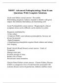
• Organizer region forms dorsal lip, blastopore, Spemann's organizer.
○ Contains/generating all these inhibitors. -> lacks all these growth factors : BMP4, Wnt8, Nodal.
○ Inhibitors secrete to the neighboring tissue. -> Ectoderm, mesoderm, endoderm.
○ NS (right) is made right above the organizer, why? It is a BMP-free zone.
○ Epidermis (right) is BMP-full.
○ Mesoderm is made from a gradient of lack of BMP (HIGH on right, LOW on left).
• By end of gastrulation and neurulation:
○ Clear A/P axis formed.
○ Notochord formed along dorsal midline.
○ Somites formation begins in anterior trunk region.
○ Lateral plate mesoderm plate established.
○ Ectoderm is induced to formmesoderm tube.
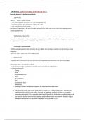
• BMP4 is a key inhibitor to neural development.
• Inhibitor is moving from organizer tissue outwards -> NS!
• Expt: cultured isolated animal cap cells (epidermis) in saline solution. -> become neural cells!
○ BMP4 is lost in the surroundings, which turns tissue back.
• ES cells does NOT get to BMP -> allowed to stay pluripotent.
• Spemann's organizer
• Expt: organizer tissue transferred to an early gastrula -> embryo developed 2 dorsal sides (two heads).
• Henson's Node transplantation
• Amphibian expt's can be done in birds.
• Wnts and Retinoic acid.
• Mesoderm tails ectoderm. Since mesoderm contains all these inhibitors -> makes them become anterior
neuro-ectoderm (head)! Chordin, Noggin, Cerberus used all up here…
• Signals Wnt and retinoic acid induce posterior ectoderm.
• Event occuring after gastrulation:
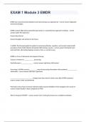
1. Somite formation
• Mesoderm paired-blocks that surround the notochord. Give rise to vertebrae and rib, bone/cartilage of
trunk, skeletal muscle, dermis of skin.
• Somites surrounding the notochord, below the neural tube, on dorsal side of embryo.
• Chick mesoderm form an anterior to the regressing node.
• Somites give rise to cervical, thorax, lumbo-sacral region.
• Somitogenic stem cells come from Henson's node.
• The temporal order of somite formation.
• Pre-somitic mesoderm does not change somite formation when transplanted/rotated. -> follow original
tissue plan. Pair of ribs.
• Yet when pre-somitic posterior mesoderm is placed into the cervical region of another chick embryo, a
pair of ribs formed in neck!
• Posterior half of somite becomes sclerotome -> cartilage of vertebrae.
• Sclerotome wraps around neural tube -> NS become wrapped in cartilage of vertebrae.
○ Notochord -> vertebral column.
• Anterior half of somite becomes dermatome -> dermis and muscle cells.
• If part of neural tube and notochord did not form/was taken out, ventral mesoderm sends signals up ->
apoptosis occurs.
○ Signals: Shh secreted from lateral plate mesoderm and grafted notochord = sclerotome
development!
• Growth factors regulate somite patterning.
• Origin of neuro-crest cells.
• Occurs during neurulation: where neural folds fuse, neural crest cells form.
Monday, August 19, 2019 10:00 AM b/c = because
b/w = between
w/ = with
• Interaction w/ VegT and B-catenin. expt = experiment
○ B-catenin left side. ex. = example
○ VegT bottom side.
• VegT induces LOW nodal -> ventral mesoderm
• B-catenin HIGH nodal -> Organizer tissue
• Experimental evidence supporting the model.
• Organizer region forms dorsal lip, blastopore, Spemann's organizer.
○ Contains/generating all these inhibitors. -> lacks all these growth factors : BMP4, Wnt8, Nodal.
○ Inhibitors secrete to the neighboring tissue. -> Ectoderm, mesoderm, endoderm.
○ NS (right) is made right above the organizer, why? It is a BMP-free zone.
○ Epidermis (right) is BMP-full.
○ Mesoderm is made from a gradient of lack of BMP (HIGH on right, LOW on left).
• By end of gastrulation and neurulation:
○ Clear A/P axis formed.
○ Notochord formed along dorsal midline.
○ Somites formation begins in anterior trunk region.
○ Lateral plate mesoderm plate established.
○ Ectoderm is induced to formmesoderm tube.
• BMP4 is a key inhibitor to neural development.
• Inhibitor is moving from organizer tissue outwards -> NS!
• Expt: cultured isolated animal cap cells (epidermis) in saline solution. -> become neural cells!
○ BMP4 is lost in the surroundings, which turns tissue back.
• ES cells does NOT get to BMP -> allowed to stay pluripotent.
• Spemann's organizer
• Expt: organizer tissue transferred to an early gastrula -> embryo developed 2 dorsal sides (two heads).
• Henson's Node transplantation
• Amphibian expt's can be done in birds.
• Wnts and Retinoic acid.
• Mesoderm tails ectoderm. Since mesoderm contains all these inhibitors -> makes them become anterior
neuro-ectoderm (head)! Chordin, Noggin, Cerberus used all up here…
• Signals Wnt and retinoic acid induce posterior ectoderm.
• Event occuring after gastrulation:
1. Somite formation
• Mesoderm paired-blocks that surround the notochord. Give rise to vertebrae and rib, bone/cartilage of
trunk, skeletal muscle, dermis of skin.
• Somites surrounding the notochord, below the neural tube, on dorsal side of embryo.
• Chick mesoderm form an anterior to the regressing node.
• Somites give rise to cervical, thorax, lumbo-sacral region.
• Somitogenic stem cells come from Henson's node.
• The temporal order of somite formation.
• Pre-somitic mesoderm does not change somite formation when transplanted/rotated. -> follow original
tissue plan. Pair of ribs.
• Yet when pre-somitic posterior mesoderm is placed into the cervical region of another chick embryo, a
pair of ribs formed in neck!
• Posterior half of somite becomes sclerotome -> cartilage of vertebrae.
• Sclerotome wraps around neural tube -> NS become wrapped in cartilage of vertebrae.
○ Notochord -> vertebral column.
• Anterior half of somite becomes dermatome -> dermis and muscle cells.
• If part of neural tube and notochord did not form/was taken out, ventral mesoderm sends signals up ->
apoptosis occurs.
○ Signals: Shh secreted from lateral plate mesoderm and grafted notochord = sclerotome
development!
• Growth factors regulate somite patterning.
• Origin of neuro-crest cells.
• Occurs during neurulation: where neural folds fuse, neural crest cells form.