Case 5 motor movement
1. What is a reflex? Different types? How it works?
- stretch reflex: a reflex elicited by a sudden external stretching force on a muscle. the
function of the stretch reflex is to keep external forces from altering the intended
position of the body. Cannot control this
- Withdrawal reflex: not monosynaptic, the shortest route in the
withdrawal-reflex circuit involves one interneuron. Under cortex
control
- Monosynaptic and polysynaptic reflexes
- Inhibitory interneuron: inhibit motor neuron in withdrawal reflex
2. How does movement work?
Hierarchically organized
- directed by commands that cascade down through the levels of a
hierarchy —from the association cortex or the company president (the
highest levels) to the muscles or the workers (the lowest levels). The
commands that emerge from the association cortex specify general
goals rather than specific plans of action. Neither the association cortex nor the
company president routinely gets involved in the details. Parallel structure enables the
association cortex or company president to exert
control over the lower levels of the hierarchy in more
than one way.
- functional segregation: each level of the sensorimotor
and company hierarchies tends to be composed of
different units (neural structures or departments), each
of which performs a different function
- sensory feedback plays an important role in directing
the continuation of the responses that produced it. The
only responses that are not normally influenced by
sensory feedback are ballistic movements—brief, all-
or-none, high-speed movements, such as swatting a
fly.
Association cortex
- posterior parietal association cortex: directing behavior by providing spatial
information, and in directing attention. Receives input from the visual system, the
auditory system, and the somatosensory system. Output to
dorsolateral prefrontal association cortex, to the various
areas of secondary motor cortex, and to the frontal eye field.
- Dorsolateral Prefrontal Association Cortex: receives
projections from the posterior parietal cortex, and it sends
projections to areas of secondary motor cortex, to primary
motor cortex, and to the frontal eye field. related to the
response rather than to the object. The response properties of
dorsolateral prefrontal neurons suggest that decisions to
initiate voluntary movements may be made in this area of
cortex
Secondary motor cortex
- Input from association areas, output to primary motor
cortex, complex movements, often involving both sides
1
, of the body. Neurons in an area of secondary motor cortex often become more active
just prior to the initiation of a voluntary movement and continue to be active
throughout the movement. Involved in the programming of specific patterns of
movements after taking general instructions from dorsolateral prefrontal cortex
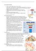
Primary motor cortex
- organized somatotopically—that is, according to a map of the body. Also called
motor homunculus
- controlling parts of the body that are capable of intricate movements, such as the
hands and mouth.
- The importance of the target of a movement, rather than the direction of a movement,
for the function of primary motor cortex
- each location in the primary motor cortex can produce innumerable patterns of muscle
contraction required to get a body part from any starting point to a target location ->
action map
- Large lesions to the primary motor cortex may disrupt a patient’s ability to move one
body part (e.g., one finger) independently of others, may produce astereognosia
(deficits in stereognosis, identifying what object you have in your hands), and may
reduce the speed, accuracy, and force of a patient’s movements. Such lesions do not,
however, eliminate voluntary movement, presumably because there are parallel
pathways that descend directly from secondary and association motor areas to
subcortical motor circuits without passing through primary motor cortex.
Cerebellum and basal ganglia
- Both the cerebellum and the basal ganglia interact with different levels of the
sensorimotor hierarchy and, in so doing, coordinate and modulate its activities.
- Cerebellum: receives information from primary and secondary motor cortex,
information about descending motor signals from
brain-stem motor nuclei, and feedback from motor
responses via the somatosensory and vestibular
systems. The cerebellum is thought to compare these
three sources of input and correct ongoing movements
that deviate from their intended course. major role in
motor learning, particularly in the learning of
sequences of movements in which timing is a critical
factor. Damage-> not controlling direction of
movement, not able to adapt to new movement
patterns, difficulty maintaining stable positions, tremor
- Basal ganglia: part of neural loops that receive cortical
input from various cortical areas and transmit it back
to the cortex via the thalamus. Modulation motor
output, cognitive functions, habit learning
Descending motor pathways
- Dorsolateral corticospinal tract: descends from the
primary motor cortex does so through the medullary
pyramids—two bulges on the ventral surface of the
medulla—then decussates and continues to descend in
the contralateral dorsolateral spinal white matter.
Direct. Wrist hands fingers toes. distal
- dorsolateral corticorubrospinal tract: descends from
the primary motor cortex synapses in the red nucleus
of the midbrain. The axons of neurons in the red
2
1. What is a reflex? Different types? How it works?
- stretch reflex: a reflex elicited by a sudden external stretching force on a muscle. the
function of the stretch reflex is to keep external forces from altering the intended
position of the body. Cannot control this
- Withdrawal reflex: not monosynaptic, the shortest route in the
withdrawal-reflex circuit involves one interneuron. Under cortex
control
- Monosynaptic and polysynaptic reflexes
- Inhibitory interneuron: inhibit motor neuron in withdrawal reflex
2. How does movement work?
Hierarchically organized
- directed by commands that cascade down through the levels of a
hierarchy —from the association cortex or the company president (the
highest levels) to the muscles or the workers (the lowest levels). The
commands that emerge from the association cortex specify general
goals rather than specific plans of action. Neither the association cortex nor the
company president routinely gets involved in the details. Parallel structure enables the
association cortex or company president to exert
control over the lower levels of the hierarchy in more
than one way.
- functional segregation: each level of the sensorimotor
and company hierarchies tends to be composed of
different units (neural structures or departments), each
of which performs a different function
- sensory feedback plays an important role in directing
the continuation of the responses that produced it. The
only responses that are not normally influenced by
sensory feedback are ballistic movements—brief, all-
or-none, high-speed movements, such as swatting a
fly.
Association cortex
- posterior parietal association cortex: directing behavior by providing spatial
information, and in directing attention. Receives input from the visual system, the
auditory system, and the somatosensory system. Output to
dorsolateral prefrontal association cortex, to the various
areas of secondary motor cortex, and to the frontal eye field.
- Dorsolateral Prefrontal Association Cortex: receives
projections from the posterior parietal cortex, and it sends
projections to areas of secondary motor cortex, to primary
motor cortex, and to the frontal eye field. related to the
response rather than to the object. The response properties of
dorsolateral prefrontal neurons suggest that decisions to
initiate voluntary movements may be made in this area of
cortex
Secondary motor cortex
- Input from association areas, output to primary motor
cortex, complex movements, often involving both sides
1
, of the body. Neurons in an area of secondary motor cortex often become more active
just prior to the initiation of a voluntary movement and continue to be active
throughout the movement. Involved in the programming of specific patterns of
movements after taking general instructions from dorsolateral prefrontal cortex
Primary motor cortex
- organized somatotopically—that is, according to a map of the body. Also called
motor homunculus
- controlling parts of the body that are capable of intricate movements, such as the
hands and mouth.
- The importance of the target of a movement, rather than the direction of a movement,
for the function of primary motor cortex
- each location in the primary motor cortex can produce innumerable patterns of muscle
contraction required to get a body part from any starting point to a target location ->
action map
- Large lesions to the primary motor cortex may disrupt a patient’s ability to move one
body part (e.g., one finger) independently of others, may produce astereognosia
(deficits in stereognosis, identifying what object you have in your hands), and may
reduce the speed, accuracy, and force of a patient’s movements. Such lesions do not,
however, eliminate voluntary movement, presumably because there are parallel
pathways that descend directly from secondary and association motor areas to
subcortical motor circuits without passing through primary motor cortex.
Cerebellum and basal ganglia
- Both the cerebellum and the basal ganglia interact with different levels of the
sensorimotor hierarchy and, in so doing, coordinate and modulate its activities.
- Cerebellum: receives information from primary and secondary motor cortex,
information about descending motor signals from
brain-stem motor nuclei, and feedback from motor
responses via the somatosensory and vestibular
systems. The cerebellum is thought to compare these
three sources of input and correct ongoing movements
that deviate from their intended course. major role in
motor learning, particularly in the learning of
sequences of movements in which timing is a critical
factor. Damage-> not controlling direction of
movement, not able to adapt to new movement
patterns, difficulty maintaining stable positions, tremor
- Basal ganglia: part of neural loops that receive cortical
input from various cortical areas and transmit it back
to the cortex via the thalamus. Modulation motor
output, cognitive functions, habit learning
Descending motor pathways
- Dorsolateral corticospinal tract: descends from the
primary motor cortex does so through the medullary
pyramids—two bulges on the ventral surface of the
medulla—then decussates and continues to descend in
the contralateral dorsolateral spinal white matter.
Direct. Wrist hands fingers toes. distal
- dorsolateral corticorubrospinal tract: descends from
the primary motor cortex synapses in the red nucleus
of the midbrain. The axons of neurons in the red
2