Case 4 brain scans
Dti = diffusion tensor imaging
Ct = computer tomography
Human optical technique
Structural imaging and functioning imaging, resolution, invasiveness
High temporal resolution-> low spatial resolution
High spatial resolution -> low temporal resolution
1. All methods categorize by temporal, spatial and functional resolution, and
invasiveness
x-ray based techniques
- x-ray photograph is taken, an x-ray beam is passed through an object and then onto a
photographic plate. Each of the molecules through which the beam passes absorbs
some of the radiation; thus, only the unabsorbed portions of the beam reach the
photographic plate. X-ray photography is therefore effective in characterizing internal
structures that differ substantially from their surroundings in the degree to which they
absorb x-rays
- contrast X-rays: Contrast x-ray techniques involve injecting into one compartment
of the body a substance that absorbs x-rays either less than or more than the
surrounding tissue. The injected substance then heightens the contrast between the
compartment and the surrounding tissue during x-ray photography.
- Computed tomography (CT/CIT): computer-assisted x-ray procedure that can be used
to visualize the brain and other internal structures of the living body. On one side of
the cylinder is an x-ray tube that projects an x-ray beam through the head to an x-ray
detector mounted on the other side. The x-ray tube and detector automatically rotate
around the head of the patient at one level of the brain, taking many individual xray
photographs as they rotate. The meager information in each x-ray photograph is
combined by a computer to generate a CT scan of one horizontal section of the brain.
Then the x-ray tube and detector are moved along the axis of the patient’s body to
another level of the brain, and the process is repeated. Scans of eight or nine horizontal
brain sections are typically obtained from a patient; when combined, they can provide
three-dimensional representations of the brain. To detect tumors and abnormalities.
Non-invasive, medium temporal and spatial resolution, no functional resolution
Radioactivity based techniques
- Positron emission tomography: radioactive fluorodeoxyglucose (FDG) is injected
into the patient’s carotid artery (an artery of the neck that feeds the ipsilateral cerebral
hemisphere). Because of its similarity to glucose, the primary metabolic fuel of the
brain, fluorodeoxyglucose is rapidly taken up by active (energy-consuming) cells.
Each PET scan is an image of the levels of radioactivity (indicated by color coding) in
various parts of one horizontal level of the brain. PET scans are not really images of
the brain. Each PET scan is merely a colored map of the amount of radioactivity in
each of the tiny cubic voxels (volume pixels) that compose the scan. The most
significant current application of PET technology is its use in identifying the
distribution in the brain of molecules of interest. Invasive, medium spatial resolution,
relative poor temporal resolution
Magnetic field based techniques
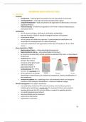
- Magnetic resonance imaging (MRI): structural brain-imaging procedure in which
high-resolution images are constructed from the measurement of radio-frequency
waves that hydrogen atoms emit as they align with a powerful magnetic field. MRI
1
Dti = diffusion tensor imaging
Ct = computer tomography
Human optical technique
Structural imaging and functioning imaging, resolution, invasiveness
High temporal resolution-> low spatial resolution
High spatial resolution -> low temporal resolution
1. All methods categorize by temporal, spatial and functional resolution, and
invasiveness
x-ray based techniques
- x-ray photograph is taken, an x-ray beam is passed through an object and then onto a
photographic plate. Each of the molecules through which the beam passes absorbs
some of the radiation; thus, only the unabsorbed portions of the beam reach the
photographic plate. X-ray photography is therefore effective in characterizing internal
structures that differ substantially from their surroundings in the degree to which they
absorb x-rays
- contrast X-rays: Contrast x-ray techniques involve injecting into one compartment
of the body a substance that absorbs x-rays either less than or more than the
surrounding tissue. The injected substance then heightens the contrast between the
compartment and the surrounding tissue during x-ray photography.
- Computed tomography (CT/CIT): computer-assisted x-ray procedure that can be used
to visualize the brain and other internal structures of the living body. On one side of
the cylinder is an x-ray tube that projects an x-ray beam through the head to an x-ray
detector mounted on the other side. The x-ray tube and detector automatically rotate
around the head of the patient at one level of the brain, taking many individual xray
photographs as they rotate. The meager information in each x-ray photograph is
combined by a computer to generate a CT scan of one horizontal section of the brain.
Then the x-ray tube and detector are moved along the axis of the patient’s body to
another level of the brain, and the process is repeated. Scans of eight or nine horizontal
brain sections are typically obtained from a patient; when combined, they can provide
three-dimensional representations of the brain. To detect tumors and abnormalities.
Non-invasive, medium temporal and spatial resolution, no functional resolution
Radioactivity based techniques
- Positron emission tomography: radioactive fluorodeoxyglucose (FDG) is injected
into the patient’s carotid artery (an artery of the neck that feeds the ipsilateral cerebral
hemisphere). Because of its similarity to glucose, the primary metabolic fuel of the
brain, fluorodeoxyglucose is rapidly taken up by active (energy-consuming) cells.
Each PET scan is an image of the levels of radioactivity (indicated by color coding) in
various parts of one horizontal level of the brain. PET scans are not really images of
the brain. Each PET scan is merely a colored map of the amount of radioactivity in
each of the tiny cubic voxels (volume pixels) that compose the scan. The most
significant current application of PET technology is its use in identifying the
distribution in the brain of molecules of interest. Invasive, medium spatial resolution,
relative poor temporal resolution
Magnetic field based techniques
- Magnetic resonance imaging (MRI): structural brain-imaging procedure in which
high-resolution images are constructed from the measurement of radio-frequency
waves that hydrogen atoms emit as they align with a powerful magnetic field. MRI
1