Lecture 6 the gastrointestinal tract
- Disease: have to compare to normal situation
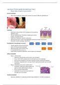
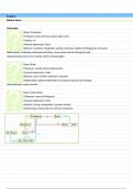
Normal esophagus
- Pink: wall of esophagus: darker pink: insertion of stomach, different epithelial layer
Esophagus
- Endoscopy: look at inside of the esophagus at the squamous
epithelium layer
- Mucosa: epithelial layer, fibroblasts, lymphocytes, lamina
propria, muscularis mucosa
- Fatty tissue: fibroblasts, vascular: submucosa,
- Thick muscular layer, circular and longitudinal: formed by smooth muscle layers
- Adventitia: outside esophagus: fatty tissue
Development of oesophageal carcinoma
- Usually sequence of precursors known
- Starts with long during oesophagitis
- Adenocarcinoma: most common, how
is it possible that these occur in a
organ lined by squamous epithelium: can be replaces by metaplasia
Reflux oesophagitis
- Phase 1: Inflammation
o Hyperemia, granulocytes and in severe cases ulceration
o Occurs when acidic content of the stomach goes up to the esophagus
o Not necessary dangerous: if it keeps on going can lead to intestinal metaplasia
o Metaplasia: important term: one type of cell is replaced by another type normally
not present
- Phase 2: Metaplasia and (chronic) inflammation
o metaplasia: replacement differentiated cell
type by another differentiated cell type
o Due to constant inflammation
o Called this: because these tissue types are
normally seen in the bowel
Intestinal metaplasia
- Squamous epithelium is replaced by intestinal type epithelium (= intestinal metaplasia)
,Dysplasia:
- on the long term if inflammation continues
- Cytonuclear atypia
o Large nuclei
o Irregular shape of nuclei
o Coarse chromatin pattern
o No lining visible
o Cytoplasm reduced
o Increases the risk of developing adenocarcinoma
Adenocarcinoma of the oesophagus (=Barrett carcinoma)
- Tumor will infiltrate into the mucosa and further
- Main difference dysplasia and carcinoma:
o Dysplasia: cells have mutation but cells still located in the are they are supposed to, if
they start invading: carcinoma created
squamous cell carcinoma
Dysplasia of squamous epithelium:
- similar to precursor carcinoma
- Normal: straight line between epithelial and lamina propria
- Dysplasia; irregular shaped cells, bigger and no differentiation between cells, still sharp
contrast between epithelial and lamina: no invasion: only dysplasia
Squamous cell carcinoma
- Tumor cells infiltrate deeper cells
stomach
Gastric mucosa
- more or less the same layers as esophagus: but epithelial cells different:
- foveolar: cells which make mucus
- lower: glandular: different types of cells
- small layer of smooth muscle: muscularis mucosa
Histology
Corpus of the stomach:
- bigger part
- Can see different types of cells in glandular layer
- 2 types
o Pink cells: parietal cells: produce acid also make intrinsic factor: binds to vitamin b12:
goes through digestive tract and will be taken up in bowels: without this factor no
uptake and results in anemia
o At the bottom more blueish: chief cells: make pepsinogen: protein important for
digestion of food: in contact with acidic stomach fluid: breaks up in pepsin molecules
which will help with digestion of food
,Antrum lower art of the stomach
- Same foveolar layer: mucus producing
- Different glandular layer: more pale, don’t find chief
or parietal cells, more mucus and antibacterial
functions
- Hormone producing cells present, difficult to
distinguish
- Gastrin: stimulates production of acidic fluid by parietal cells in corpus
Inflammation of the gastric mucosa (= gastritis)
- Acute gastritis: caused by bacteria in the stomach
o H. pylori, Alcohol, NSAID
- Auto-immune gastritis
- Others: very rare
o Granulomatous gastritis, Crohn’s disease, Lymfocytic gastritis, Other
Helicobacter pylori
- Curly cells at the surface of cells: helicobacter: pretty much only bacteria able to survive this
environment
- Can cause severe ulcers and inflammation: surgery needed, but after discovery: found out
that antibiotics can cure the inflammation and ulcers
Auto-immune gastritis
- Antibodies against parietal cells / intrinsic factor
o Can lead to severe inflammation
- Intestinal metaplasia
o Parietal cells will disappear: because cells will differentiate into intestinal type cells
- Neuro-endocrine cell hyperplasia: without parietal cells no more acid producing cells
present, these cells regulate the acidity of the stomach by activating gastrin producing cells
o Gastrin producing cells: will sense highering of pH due to less parietal cells: will start
differentiating: can lead to tumors
- Anemia: also loose intrinsic factor: needed for production of red blood cells
- Increased risk for adenocarcinoma
- Increased risk for neuro-endocrine tumor
Development of gastric adenocarcinoma
intestinal type
- not necessary in the bowel named after the cell types
- will form glandular structures: irregular shaped glands
- Has central crater
, Gastric adenocarcinoma diffuse type
- Hardly able to see the circumcised tumor area: all looks more or less the same
- The tumor is everywhere: difficult to see but the wall is thickened in all the stomach
- Thick wall which can not expand anymore: very stiff wall
- Tumor grows below the surface: difficult to see stomach cells
- Procedure repeated until tumor cell found
- Do not form glandular structure
- Invade as single cells: lost adhesion molecules
- Worse prognosis
- Both chemotherapy: both tumor types
o Diffuse type will not response as well
Colorectal cancer development
- Book: thesis inflammatory disorders
Facts about GI tumors
- GI tumors comprise 27% of all cancer related mortality
- High incidence
- Present in late stage of disease: poor prognosis
Forms of cancer prevention
- Prevention: difficult but important to promote awareness
- Etiology - Primary prevention
- Pathogenesis - Secondary prevention/Diagnostics
- Tumor biology – Therapy
Secondary preventive
- Population based screening for colorectal cancer
o Focused on 55-75
o Identification of high risk subjects
Will be focused on during screening
o Aim: Early detection or precursor detection
Logistics of the screening
- Stool tested for blood
- Positive: occult blood present: patient invited for colonoscopy
- Colonoscopy: identifies lesions in the bowel, polyps removed and looked for tumor
- Try to get the cancer before cancer development which causes symptoms
Colorectal polyps
- Cancer detected because not just atypical cells but usually form polyps: may different types
- Focused on adenoma: dysplasia in the form of a polyp present
- Prevent cancer: remove polyps
- Side effects of removing polyps: overall quite safe,
but bleeding or proliferation can occur
- Disease: have to compare to normal situation
Normal esophagus
- Pink: wall of esophagus: darker pink: insertion of stomach, different epithelial layer
Esophagus
- Endoscopy: look at inside of the esophagus at the squamous
epithelium layer
- Mucosa: epithelial layer, fibroblasts, lymphocytes, lamina
propria, muscularis mucosa
- Fatty tissue: fibroblasts, vascular: submucosa,
- Thick muscular layer, circular and longitudinal: formed by smooth muscle layers
- Adventitia: outside esophagus: fatty tissue
Development of oesophageal carcinoma
- Usually sequence of precursors known
- Starts with long during oesophagitis
- Adenocarcinoma: most common, how
is it possible that these occur in a
organ lined by squamous epithelium: can be replaces by metaplasia
Reflux oesophagitis
- Phase 1: Inflammation
o Hyperemia, granulocytes and in severe cases ulceration
o Occurs when acidic content of the stomach goes up to the esophagus
o Not necessary dangerous: if it keeps on going can lead to intestinal metaplasia
o Metaplasia: important term: one type of cell is replaced by another type normally
not present
- Phase 2: Metaplasia and (chronic) inflammation
o metaplasia: replacement differentiated cell
type by another differentiated cell type
o Due to constant inflammation
o Called this: because these tissue types are
normally seen in the bowel
Intestinal metaplasia
- Squamous epithelium is replaced by intestinal type epithelium (= intestinal metaplasia)
,Dysplasia:
- on the long term if inflammation continues
- Cytonuclear atypia
o Large nuclei
o Irregular shape of nuclei
o Coarse chromatin pattern
o No lining visible
o Cytoplasm reduced
o Increases the risk of developing adenocarcinoma
Adenocarcinoma of the oesophagus (=Barrett carcinoma)
- Tumor will infiltrate into the mucosa and further
- Main difference dysplasia and carcinoma:
o Dysplasia: cells have mutation but cells still located in the are they are supposed to, if
they start invading: carcinoma created
squamous cell carcinoma
Dysplasia of squamous epithelium:
- similar to precursor carcinoma
- Normal: straight line between epithelial and lamina propria
- Dysplasia; irregular shaped cells, bigger and no differentiation between cells, still sharp
contrast between epithelial and lamina: no invasion: only dysplasia
Squamous cell carcinoma
- Tumor cells infiltrate deeper cells
stomach
Gastric mucosa
- more or less the same layers as esophagus: but epithelial cells different:
- foveolar: cells which make mucus
- lower: glandular: different types of cells
- small layer of smooth muscle: muscularis mucosa
Histology
Corpus of the stomach:
- bigger part
- Can see different types of cells in glandular layer
- 2 types
o Pink cells: parietal cells: produce acid also make intrinsic factor: binds to vitamin b12:
goes through digestive tract and will be taken up in bowels: without this factor no
uptake and results in anemia
o At the bottom more blueish: chief cells: make pepsinogen: protein important for
digestion of food: in contact with acidic stomach fluid: breaks up in pepsin molecules
which will help with digestion of food
,Antrum lower art of the stomach
- Same foveolar layer: mucus producing
- Different glandular layer: more pale, don’t find chief
or parietal cells, more mucus and antibacterial
functions
- Hormone producing cells present, difficult to
distinguish
- Gastrin: stimulates production of acidic fluid by parietal cells in corpus
Inflammation of the gastric mucosa (= gastritis)
- Acute gastritis: caused by bacteria in the stomach
o H. pylori, Alcohol, NSAID
- Auto-immune gastritis
- Others: very rare
o Granulomatous gastritis, Crohn’s disease, Lymfocytic gastritis, Other
Helicobacter pylori
- Curly cells at the surface of cells: helicobacter: pretty much only bacteria able to survive this
environment
- Can cause severe ulcers and inflammation: surgery needed, but after discovery: found out
that antibiotics can cure the inflammation and ulcers
Auto-immune gastritis
- Antibodies against parietal cells / intrinsic factor
o Can lead to severe inflammation
- Intestinal metaplasia
o Parietal cells will disappear: because cells will differentiate into intestinal type cells
- Neuro-endocrine cell hyperplasia: without parietal cells no more acid producing cells
present, these cells regulate the acidity of the stomach by activating gastrin producing cells
o Gastrin producing cells: will sense highering of pH due to less parietal cells: will start
differentiating: can lead to tumors
- Anemia: also loose intrinsic factor: needed for production of red blood cells
- Increased risk for adenocarcinoma
- Increased risk for neuro-endocrine tumor
Development of gastric adenocarcinoma
intestinal type
- not necessary in the bowel named after the cell types
- will form glandular structures: irregular shaped glands
- Has central crater
, Gastric adenocarcinoma diffuse type
- Hardly able to see the circumcised tumor area: all looks more or less the same
- The tumor is everywhere: difficult to see but the wall is thickened in all the stomach
- Thick wall which can not expand anymore: very stiff wall
- Tumor grows below the surface: difficult to see stomach cells
- Procedure repeated until tumor cell found
- Do not form glandular structure
- Invade as single cells: lost adhesion molecules
- Worse prognosis
- Both chemotherapy: both tumor types
o Diffuse type will not response as well
Colorectal cancer development
- Book: thesis inflammatory disorders
Facts about GI tumors
- GI tumors comprise 27% of all cancer related mortality
- High incidence
- Present in late stage of disease: poor prognosis
Forms of cancer prevention
- Prevention: difficult but important to promote awareness
- Etiology - Primary prevention
- Pathogenesis - Secondary prevention/Diagnostics
- Tumor biology – Therapy
Secondary preventive
- Population based screening for colorectal cancer
o Focused on 55-75
o Identification of high risk subjects
Will be focused on during screening
o Aim: Early detection or precursor detection
Logistics of the screening
- Stool tested for blood
- Positive: occult blood present: patient invited for colonoscopy
- Colonoscopy: identifies lesions in the bowel, polyps removed and looked for tumor
- Try to get the cancer before cancer development which causes symptoms
Colorectal polyps
- Cancer detected because not just atypical cells but usually form polyps: may different types
- Focused on adenoma: dysplasia in the form of a polyp present
- Prevent cancer: remove polyps
- Side effects of removing polyps: overall quite safe,
but bleeding or proliferation can occur