BSCI202 lab blood vessels
General circulation
▪ Closed transport system
▪ Heart → large arteries → medium-sized arteries → arterioles → capillary beds →
venules →medium-sized veins → large veins → heart
▪ Substances diffuse across capillary walls
Microscopic structure (page 364)
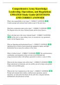
▪ 3 tunics surrounding the lumen:
o Tunica intima/ interna
- Innermost layer
- Thin layer of endothelium w/ CT basement membrane
- Continuation of endocardium and with the ventricles in the atria
- Slick surface
o Tunica media
- Circularly arranged smooth muscle and elastic CT
- Allows for changes in lumen diameter
- Lots of smooth muscle & elastic (connective) tissue (because needs strength
and flexibility)
- This layer is not as thick in the venous system as the arteries system
o Tunica adventitia/ externa
- Most superficial
- Areolar or fibrous CT
- Protects and anchors vessels
- Nerve fibers, lymph vessels, blood vessels
only thing left in the capillary bed is the tunica interna (one
layer of endothelium tissue)
know that the left side is the tunica media because of the
muscular wall in the artery (even though there is no blood
the artery will hold its shape while a vein would collapse)
, vein is mostly 2x as large as arteries
Vessel Types (page 365)
▪ Arteries
o Thicker, more structural integrity because under higher pressure
- Under the greatest systemic pressure
o Transport blood away from heart
o Expands during systole, recoils passively during diastole (can be oxygen rich or
not)
o Types of arteries
- Elastic arteries: close to heart, large, “conducting vessels”, most expandable
the aorta
- Muscular arteries: medium-sized, “distributing vessels”, named arteries in the
body transporting vessels
- Arterioles: smallest, “resistance vessels” go into the capillary bed
▪ Capillaries
o narrowest vessels
o Walls = endothelium (one cell layer)
o Involved in gas, nutrient, & waste exchange
▪ Veins
o Transport blood toward the heart
o 65% of blood volume is in veins – Capacitance vessels
o Low pressure vessels because of thin walls and large lumens
o Adaptations for venous return blood to Right Atrium:
- Valves: formed by folds of tunica intima
- prevent back flow
- highly concentrated in limbs
Veins
▪ Adaptations for venous return = cardiac output:
o Skeletal muscle pump: Skeletal muscle contractions squeeze veins (Vascular
regulation) and helps prepull blood up
- And valves close to prevent back flow
- Helps getting blood to the thoracic region
o Pressure from blood coming from capillary beds
- Pushes blood up each segment and can’t go back to the previous one because
there are valves
- Way to get blood back to the heart because the venous system is under low
pressure
▪ Respiratory pump
o During inspiration, a decrease in intra -thoracic pressure (diaphragm will move
down) and increase intra -abdominal pressure causes blood flow from veins in the
abdominal region to veins in the thoracic region
- Creates a vacuum which brings the venous blood up to the heart
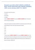
Major arteries of system circulation
, ▪ Aorta
o Largest artery in the body
o Leaves from the left ventricle of the heart
o Regions
- Ascending aorta - leaves the left ventricle
- Aortic arch - arches to the left
- Thoracic aorta - travels downward through the thorax (t5 to t12)
- Abdominal aorta - passes through the diaphragm into the abdominopelvic
cavity
- Last two are part of the descending aorta
Page 368
▪ Arterial branches of the ascending aorta
o Right and left coronary arteries serve the heart
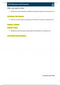
▪ Arterial branches of the aortic arch
o Brachiocephalic trunk splits into the
- Right common carotid artery
- Right subclavian artery
o Left common carotid artery splits into the
- Left internal and external carotid arteries
o Left subclavian artery branches into the
- Vertebral artery
- In the axilla, the subclavian artery becomes the axillary artery brachial
artery radial and ulnar arteries
Page 369
▪ Arterial branches of the thoracic aorta
o Intercostal arteries supply the muscles of the thorax wall
o Other branches of the thoracic aorta supply the
- Lungs (bronchial arteries)
- Esophagus (esophageal arteries)
- Diaphragm (phrenic arteries)
Page 371
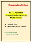
▪ Arterial branches of the abdominal aorta
o Celiac trunk is the first branch of the abdominal aorta.
- Three branches are
- Left gastric artery (stomach)
- Splenic artery (spleen)
- Common hepatic artery (liver)
o Superior mesenteric artery supplies most of the small intestine and first half of the
large intestine
o Left and right renal arteries (kidney)
o Left and right gonadal arteries
- Ovarian arteries in females serve the ovaries
- Testicular arteries in males serve the testes
General circulation
▪ Closed transport system
▪ Heart → large arteries → medium-sized arteries → arterioles → capillary beds →
venules →medium-sized veins → large veins → heart
▪ Substances diffuse across capillary walls
Microscopic structure (page 364)
▪ 3 tunics surrounding the lumen:
o Tunica intima/ interna
- Innermost layer
- Thin layer of endothelium w/ CT basement membrane
- Continuation of endocardium and with the ventricles in the atria
- Slick surface
o Tunica media
- Circularly arranged smooth muscle and elastic CT
- Allows for changes in lumen diameter
- Lots of smooth muscle & elastic (connective) tissue (because needs strength
and flexibility)
- This layer is not as thick in the venous system as the arteries system
o Tunica adventitia/ externa
- Most superficial
- Areolar or fibrous CT
- Protects and anchors vessels
- Nerve fibers, lymph vessels, blood vessels
only thing left in the capillary bed is the tunica interna (one
layer of endothelium tissue)
know that the left side is the tunica media because of the
muscular wall in the artery (even though there is no blood
the artery will hold its shape while a vein would collapse)
, vein is mostly 2x as large as arteries
Vessel Types (page 365)
▪ Arteries
o Thicker, more structural integrity because under higher pressure
- Under the greatest systemic pressure
o Transport blood away from heart
o Expands during systole, recoils passively during diastole (can be oxygen rich or
not)
o Types of arteries
- Elastic arteries: close to heart, large, “conducting vessels”, most expandable
the aorta
- Muscular arteries: medium-sized, “distributing vessels”, named arteries in the
body transporting vessels
- Arterioles: smallest, “resistance vessels” go into the capillary bed
▪ Capillaries
o narrowest vessels
o Walls = endothelium (one cell layer)
o Involved in gas, nutrient, & waste exchange
▪ Veins
o Transport blood toward the heart
o 65% of blood volume is in veins – Capacitance vessels
o Low pressure vessels because of thin walls and large lumens
o Adaptations for venous return blood to Right Atrium:
- Valves: formed by folds of tunica intima
- prevent back flow
- highly concentrated in limbs
Veins
▪ Adaptations for venous return = cardiac output:
o Skeletal muscle pump: Skeletal muscle contractions squeeze veins (Vascular
regulation) and helps prepull blood up
- And valves close to prevent back flow
- Helps getting blood to the thoracic region
o Pressure from blood coming from capillary beds
- Pushes blood up each segment and can’t go back to the previous one because
there are valves
- Way to get blood back to the heart because the venous system is under low
pressure
▪ Respiratory pump
o During inspiration, a decrease in intra -thoracic pressure (diaphragm will move
down) and increase intra -abdominal pressure causes blood flow from veins in the
abdominal region to veins in the thoracic region
- Creates a vacuum which brings the venous blood up to the heart
Major arteries of system circulation
, ▪ Aorta
o Largest artery in the body
o Leaves from the left ventricle of the heart
o Regions
- Ascending aorta - leaves the left ventricle
- Aortic arch - arches to the left
- Thoracic aorta - travels downward through the thorax (t5 to t12)
- Abdominal aorta - passes through the diaphragm into the abdominopelvic
cavity
- Last two are part of the descending aorta
Page 368
▪ Arterial branches of the ascending aorta
o Right and left coronary arteries serve the heart
▪ Arterial branches of the aortic arch
o Brachiocephalic trunk splits into the
- Right common carotid artery
- Right subclavian artery
o Left common carotid artery splits into the
- Left internal and external carotid arteries
o Left subclavian artery branches into the
- Vertebral artery
- In the axilla, the subclavian artery becomes the axillary artery brachial
artery radial and ulnar arteries
Page 369
▪ Arterial branches of the thoracic aorta
o Intercostal arteries supply the muscles of the thorax wall
o Other branches of the thoracic aorta supply the
- Lungs (bronchial arteries)
- Esophagus (esophageal arteries)
- Diaphragm (phrenic arteries)
Page 371
▪ Arterial branches of the abdominal aorta
o Celiac trunk is the first branch of the abdominal aorta.
- Three branches are
- Left gastric artery (stomach)
- Splenic artery (spleen)
- Common hepatic artery (liver)
o Superior mesenteric artery supplies most of the small intestine and first half of the
large intestine
o Left and right renal arteries (kidney)
o Left and right gonadal arteries
- Ovarian arteries in females serve the ovaries
- Testicular arteries in males serve the testes