H13: EYE DEVELOPMENT
ORGANOGENESIS
P. CALLAERTS
Inhoudsopgave
13.1 THE ADULT MAMMALIAN EYE.............................................................................3
13.1.1 THE ADULT MAMMALIAN RETINA................................................................................................3
13.2 THE ADULT DROSOPHILA EYE.............................................................................4
13.3 EYE DEVELOPMENT IN MAMMALS.......................................................................5
13.3.1 EYE DEVELOPMENT IN MAMMALS: EMBRYONIC LINEAGES.................................................................6
13.4 EYE DEVELOPMENT IN DROSOPHILA...................................................................6
13.5 EYE DEVELOPMENT: INSPIRATIONAL EXPERIMENTS.............................................7
13.5.1 REMOVAL OF EYE VESICLE........................................................................................................ 7
13.5.2 TRANSPLANTATION OF OPTIC CUP.............................................................................................. 8
13.6 EYE DEVELOPMENT: DECODE MOLECULAR MECHANISMS.....................................8
13.6.1 PAX 6 IN MAMMALIAN EYE DEVELOPMENT..................................................................................10
13.6.2 PAX 6 IN OTHER TISSUES....................................................................................................... 10
13.7 EYE DEVELOPMENT DROSOPHILA: DECODE MOLECULAR MECHANISMS -EYELESS,
TWIN OF EYELESS, SINE OCULIS, EYES ABSENT.........................................................11
13.8 EYE DEVELOPMENT: ANALYSIS OF GENE FUNCTION – OVEREXPRESSION STUDIES
.............................................................................................................................. 11
13.9 EYE DEVELOPMENT DROSOPHILA: GENE NETWORKS..........................................12
13.10 EYE DEVELOPMENT MAMMALS: GENE NETWORKS............................................14
13.11 EYE DEVELOPMENT IN MAMMALS: AXES IN THE OPTIC CUP..............................15
13.12 EYE DEVELOPMENT IN MAMMALS: TEMPORAL PROGRESSION OF RETINOGENESIS
IN THE MOUSE........................................................................................................ 15
13.13 EYE DEVELOPMENT IN MAMMALS: RETINAL CELL FATES...................................16
13.14 IMPLICATIONS IN BIOMEDICINE – CONGENITAL EYE DEFECTS............................16
13.14.1 OTX2 MUTATIONS............................................................................................................. 17
13.14.2 SOX2 MUTATIONS............................................................................................................. 17
13.14.3 RAX MUTATIONS............................................................................................................... 17
13.14.4 CRX MUTATIONS................................................................................................................. 18
13.14.5 ATONAL HOMOLOG 7 (ATOH7) LOSS OF FUNCTION MUTATIONS...................................................19
1
,13.15 IMPLICATIONS IN BIOMEDICINE – UNDERSTANDING TERATOGENS.....................19
Goals:
To know the basic structure of the adult mammalian and Drosophila eye
To distinguish key steps in mammalian eye development + to identify contributing
embryonic lineages.
To know the organization of the Drosophila eye-antennal disc
To be able to interpret Hans Spemann’s experiments related to the eye.
To know the structure and function of Pax6 in the eye of different animals
To know and be able to interpret key experiments in Drosophila that resulted in the
first framework for the genetic control of early eye development and discuss their
relevance for mammalian eye development.
To know key implications in biomedicine of eye development associated genes and be
able to identify which developmental step is likely interrupted.
EXAM:
Picture of either drosophila or mammalian eye and you have to identify the different
components. An arrow is pointing to a structure what structure is that?
Able to identify key steps in mammalian eye development and identify what embryonic
lineages contribute to that.
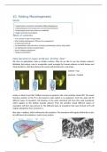
Vision: solving a problem in different ways
If you are an organism that can give light, that will give you some selective advantage but there
is also some sort of pigment cells present. Wherever the lights come from, it allows orientation
movement. Either to the light or away of the light and so that innovation is what we see in a
broad range of species.
2
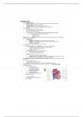
, 13.1 THE ADULT MAMMALIAN EYE
Overview of adult mouse eye Overview of adult human
eye
Overview:
- The lens (the refractory organ)
- Anterior from it: pupil that is covered by the cornea.
- Around: iris (part that is colored and contracts). This contraction comes from the muscles
in the ciliary body. Little muscles that can contract or relax dependent on which light the
eye is exposed.
- The sclera is the hard layer around the eye.
- In the back of the eye is the retina. That is the photosensitive part and all the
photoreceptors they send their info to the brain through the optic nerve.
Between the retina and the sclera is the choroid. The choroid is the blood vessels rich
area that provides nutrition and oxygen to the retinal cells.
In the human eye: we have the macula lutea (centrally positioned to the lens) and the fovea
centralis (rich in cone photoreceptors). 2 typed: rod and cone photoreceptors. Cone receptors are
in a very high density positioned central in the eye so we can have very precise and specific
vision.
13.1.1 THE ADULT MAMMALIAN RETINA
RPE: retinal pigmented
epithelium
OS: outer segment
IS: inner segment
ONL: outer nuclear layer
OPL: outer plexiform layer
INL: inner nuclear layer
IPL: inner plexiform layer
3 RGCL: retinal ganglion cell layer
ORGANOGENESIS
P. CALLAERTS
Inhoudsopgave
13.1 THE ADULT MAMMALIAN EYE.............................................................................3
13.1.1 THE ADULT MAMMALIAN RETINA................................................................................................3
13.2 THE ADULT DROSOPHILA EYE.............................................................................4
13.3 EYE DEVELOPMENT IN MAMMALS.......................................................................5
13.3.1 EYE DEVELOPMENT IN MAMMALS: EMBRYONIC LINEAGES.................................................................6
13.4 EYE DEVELOPMENT IN DROSOPHILA...................................................................6
13.5 EYE DEVELOPMENT: INSPIRATIONAL EXPERIMENTS.............................................7
13.5.1 REMOVAL OF EYE VESICLE........................................................................................................ 7
13.5.2 TRANSPLANTATION OF OPTIC CUP.............................................................................................. 8
13.6 EYE DEVELOPMENT: DECODE MOLECULAR MECHANISMS.....................................8
13.6.1 PAX 6 IN MAMMALIAN EYE DEVELOPMENT..................................................................................10
13.6.2 PAX 6 IN OTHER TISSUES....................................................................................................... 10
13.7 EYE DEVELOPMENT DROSOPHILA: DECODE MOLECULAR MECHANISMS -EYELESS,
TWIN OF EYELESS, SINE OCULIS, EYES ABSENT.........................................................11
13.8 EYE DEVELOPMENT: ANALYSIS OF GENE FUNCTION – OVEREXPRESSION STUDIES
.............................................................................................................................. 11
13.9 EYE DEVELOPMENT DROSOPHILA: GENE NETWORKS..........................................12
13.10 EYE DEVELOPMENT MAMMALS: GENE NETWORKS............................................14
13.11 EYE DEVELOPMENT IN MAMMALS: AXES IN THE OPTIC CUP..............................15
13.12 EYE DEVELOPMENT IN MAMMALS: TEMPORAL PROGRESSION OF RETINOGENESIS
IN THE MOUSE........................................................................................................ 15
13.13 EYE DEVELOPMENT IN MAMMALS: RETINAL CELL FATES...................................16
13.14 IMPLICATIONS IN BIOMEDICINE – CONGENITAL EYE DEFECTS............................16
13.14.1 OTX2 MUTATIONS............................................................................................................. 17
13.14.2 SOX2 MUTATIONS............................................................................................................. 17
13.14.3 RAX MUTATIONS............................................................................................................... 17
13.14.4 CRX MUTATIONS................................................................................................................. 18
13.14.5 ATONAL HOMOLOG 7 (ATOH7) LOSS OF FUNCTION MUTATIONS...................................................19
1
,13.15 IMPLICATIONS IN BIOMEDICINE – UNDERSTANDING TERATOGENS.....................19
Goals:
To know the basic structure of the adult mammalian and Drosophila eye
To distinguish key steps in mammalian eye development + to identify contributing
embryonic lineages.
To know the organization of the Drosophila eye-antennal disc
To be able to interpret Hans Spemann’s experiments related to the eye.
To know the structure and function of Pax6 in the eye of different animals
To know and be able to interpret key experiments in Drosophila that resulted in the
first framework for the genetic control of early eye development and discuss their
relevance for mammalian eye development.
To know key implications in biomedicine of eye development associated genes and be
able to identify which developmental step is likely interrupted.
EXAM:
Picture of either drosophila or mammalian eye and you have to identify the different
components. An arrow is pointing to a structure what structure is that?
Able to identify key steps in mammalian eye development and identify what embryonic
lineages contribute to that.
Vision: solving a problem in different ways
If you are an organism that can give light, that will give you some selective advantage but there
is also some sort of pigment cells present. Wherever the lights come from, it allows orientation
movement. Either to the light or away of the light and so that innovation is what we see in a
broad range of species.
2
, 13.1 THE ADULT MAMMALIAN EYE
Overview of adult mouse eye Overview of adult human
eye
Overview:
- The lens (the refractory organ)
- Anterior from it: pupil that is covered by the cornea.
- Around: iris (part that is colored and contracts). This contraction comes from the muscles
in the ciliary body. Little muscles that can contract or relax dependent on which light the
eye is exposed.
- The sclera is the hard layer around the eye.
- In the back of the eye is the retina. That is the photosensitive part and all the
photoreceptors they send their info to the brain through the optic nerve.
Between the retina and the sclera is the choroid. The choroid is the blood vessels rich
area that provides nutrition and oxygen to the retinal cells.
In the human eye: we have the macula lutea (centrally positioned to the lens) and the fovea
centralis (rich in cone photoreceptors). 2 typed: rod and cone photoreceptors. Cone receptors are
in a very high density positioned central in the eye so we can have very precise and specific
vision.
13.1.1 THE ADULT MAMMALIAN RETINA
RPE: retinal pigmented
epithelium
OS: outer segment
IS: inner segment
ONL: outer nuclear layer
OPL: outer plexiform layer
INL: inner nuclear layer
IPL: inner plexiform layer
3 RGCL: retinal ganglion cell layer