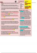
,CASE 3
, anatomy
coronary
stabilises3
vessels
·
anchors
subsections ·
limits expan-
Sion arteries
)
& position
pericardium
-
① anteor
pleural fibrous outer 3 thickest marginal
RCA
·
between around
cavities layer circumfRx goes
a LCA
in mediastinum parietal lines fibrous
Spericardi to posterior
·
anterior interventricular
·
·
1. aveolar
·
pericardial Jac 2 Mesothelim
. PC fluid
attached to
· anteroposteriorly · visceral 2 posterior LCA
from
·
posterior to sternum
heart wall / circum flex
↳ marginal
2
3rd C
·
base :
·
epicardium outer
to posterior - marginal
: 5th 1C space same structure RCA wraps
apex
·
as visceral PC
mesothelium
+
a reolar Ct
veins
myocardium muscular
cardiac LAD
concentric layers 1
great :
· endocardium simple squamous
valves
2. Middle cardiac : PDA - Coronary
Small : RA , RV
3 .
O
3 atria
thin walls
: musculli pectinate
④ ventricles ↳ stronger contractions
thick walls ·
anrides
· trabeculal carnal : ·
Christa terminal is
muscular ridges ↳ crescent shaped
Arvalues chordal ridges & openings
tendinde
papillary SUC drains deoxy
RA :
musues blood from upper
tricuspid body
·
RV viscera
,
trunk , lower
moderator band : IVC :
lower limbs
part of trab corndl.
↳ shortcut in coronary : drainage point
conduction system Sinus for coronary veins
palm trunk sprm ↳ posterior a
.
semilunar value > pulm
. -
Ovalis connects atria mediastinum
fossa :
arteries before birth
bicuspid/mitral non closure ASD
superior region
:
↳
Ly I
aortic arch 3 aortic SA3AV node , internodal pathways
value borders :
·
LA pulm veins (L3Rx2)
.
thoracic
T1 -
4
F S
invet
m
& p A- manubrium
L
2 inferior ↓
pleural
Ar values
ventricle contracts a) anterior contents
-thymys
muscle between
sternum 3 -great
vessels
↳ papillary pericardium -trached + oesophagus
chordae tending
closes value
tighten =
peri-
cardium anterio
atria 3
body
I :
m
between & p A-- sternal S
ventricles
L
Si
FL diaphragm
·
↓ I :
semilunau pleun al P I : middle
3 porta
pulm trunk .
·
closed by backflow
. S2
, autorhythmic
physiology cells connected
conduction via gap junctions
·
SA node : 100 BPM
↑ musce system -
creates refractory
for other
periods
·
excitable pacemaker sites
↳ RMP depol
=
Ap AU mode
: 40-60
20-10
S
80mV
>
- -
12
plateau
+
·
k efflux = (a2 +
contractive ·
perkunje :
ii
influx Wl
discs cells
chronotropism -> affects pacemakercesp in SA
between cells
intercalated
I allows contractions
I
desmosomes
.
-
junctions in syncytium A in SA node node
-gap
regulation of HR by
ANS
-
SNS : E ,
NE = * HR contractility
: ACh inverse
PNS
:
-
cardiac APS
2
dromotropism
diff APs
different regions AV node
=
conduction through
.
rate of
↳ a ion
channel types ?
ANS
densities
atrial : short 3 flatter 3 Tonotropism
contractility ofmaa
-
M regof
needs less
+
Ca2
·
in cytoplasm
concentration
-ventricular : longer 3 Steep
↳ Cast
-
regulation
removal of
cardiac heart can adjust output irt
cazt
cytoplasmic output
curves
A in preload
↳ degree of stretch in
starling's ventricles & end of
↑ symp. did Stole
activity
Su
control
·
frank-starling :
of
-
excessive preload >
-
degree
.
symp stretch is so great =
optimal
block
mystible overlap is exceeded
- + F = ↓SV
normal
preload
CEDU)
2 stroke volume
ventricles
· amount of blood ejected from
with each contraction
·
SV : EDV-ESU
·
RV prelodd
into SR affecting factors
, reuptake ~
-
venous return
·
Ca2
+
ATPase pump (SERCA) -
diastolic filling time
atrial p
cast -
↳ actively pumps - ventricular compliance
from mysfilaments
into SR
via hydrolysis
·
contractility Staircase
phenomenon
E
ATP =
regulated by ionotropism * NB
tension
-
IT catecholamines period
of
2 extrusion from cell -
:
rest sufficient
plasma OIT : Can blockers
Na-Ca exchanger (NCX)
on HR
-
F-frequency relationship (Treppe
membrane
-
actively
Atpase uLes AtP to at
+ NCX slows down HR due to
Nat/k
wat out 3 K
+
in saturation of Na/k Atpace
more
electro accumulation of Ca2+
↳ lower Nat in cytoplasm
=
↑ F of contraction
chem gradient relaxation
summation is different of is higher
↳ drives NCX ·
is not sufficient