Pharmaceutical biotechnology
lecture notes
Introduction
Biopharmaceutical: a compound partly produced using a cell. A biopharmaceutical is:
A complex molecule
Almost always injectable
Expensive
Used for live threating diseases
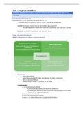
Defined biopharmaceutical Undefined biopharmaceutical
- Chemical composition/structure is - Critical product attribution unknown
known - Need extensive clinical trial testing
- Mechanism of action and side-effects - Process is not known
are known - Processes needed to produce the
- You can exactly predict the product biopharmaceutical are based on old
technologies
Most biopharmaceuticals are undefined, due to posttranslational modifications (mostly
glycosylation). Glycosylation is important for activity, immunogenicity, in vivo half-life, and stability.
Bacteria: for small proteins without glycosylation
Yeast: for complex protein structures with limited glycosylation
Animal cells: for complex protein structures with extensive glycosylation
Approval path: only 1 out of 10 products end up at the market.
1. Preclinical stage (in cells): investigate new drugs
2. Phase 1 (small group of healthy persons): investigate side-effects
3. Phase 2 (small group of patients): investigate optimal dosage and type of application
4. Phase 3 (large group of patients): investigate effectiveness and final safety
To go through all the phases, the costs are around 2 billion euros.
Fast track: for orphan drugs (disease with very few patients) and public health advantages (covid).
The drug is immediately applied to patients, the large trials are skipped.
Biosimilars: copying of an off patent biopharmaceutical, less trials needed.
Defined products also need less clinical trials.
Animal cell cultures can be used for:
In vitro studies (for basic cell physiology, toxicity, drug candidates, and food components)
Cell therapy
Tissue engineering (in vitro hamburger)
Pharmaceutical proteins (EPO) production
Viral vaccines production
Hayflick limit: there is a limit to the amount of division each cell can take.
Some cells can escape this Hayflick limit (see image), and these cells include
,stem cells and some transformed cells (for example cancer cells). The transformed cells are used in
biotechnology. Transformed cells characteristics:
Infinite life span
Acceptable growth rate
Low growth factor dependence
Suspension growth
Aneuploid (a mutation where more or less chromosomes occur)
Mutagens, viruses, and oncogenes can be used to produce transformed cells. Transformation
sometimes happened spontaneously. You can also take cells from tumours to use them for
applications.
If you fill a bioreactor with E.coli, it will grow and fill the entire bioreactor (maximum cell density). For
insect and mammalian cells, you need a minimal cell density. If the density is too low, the cells will
not start growing. The maximum cell density of animal cells is also much lower than for E.coli cells (in
a normal batch). The medium for animal cells is very complex, making the medium very expensive as
well. The media are very rich: everything is present. This means that if the medium is infected with an
E.coli cell or another bacterium, the bacterium will outgrow your cell culture. Cell contamination is
the major concern.
You can add serum to your medium. The functions and problems of
serum are shown in the image.
Shear sensitivity is a problem for scale-up. Shear sensitivity is caused
by a lack of cell walls and a decrease in cell size.
A comparison:
Mammalian cells Insect cells Yeast cells Bacterial cells
Nutrition Complex Complex Simple Simple
Media costs 10-100 10-100 <1 <1
(euro per dm3)
Secretion Yes Yes/lytic Variable No
Posttranslational ++ + +/- --
modifications
Downstream Simple Simple Complex Complex
processing
Scale-up Difficult Difficult Simple Simple
Cell proliferation and death
Cell death decreases the amount of viable cells and results in the release of cell contents. Cell
contents include:
Proteases
Unprocessed product
Host cell proteins
DNA
Cell death decreases product quality and product yield.
When cells are in a quiescent state, you can get the cells into the competent stage against. Thus, a
differentiated cell can divide again. In order to achieve this, you must change some physiological
stages, for example the growth factors. Quiescent cells can also go into apoptosis. Cells can be in
, division, in quiescent cells or they can be death cells (due to apoptosis and necrosis).
The cancer cells do not go into quiescent stages: they either divide or they die.
Acridine: can stain DNA (gives a green spot) -> early apoptotic stage.
PI: can stain DNA as well but gives a much stronger staining (gives a red
spot). PI only gives a staining when the membrane has become permeable
-> late apoptotic stage.
Acridine and PI can be used to track apoptosis and necrosis in cells. With
apoptosis, stress factors are slowly added to break the cytoskeleton. Water
is excreted from the cell to condensate the DNA.
Necrosis happens in response to a very high level of stress.
The cell has a positively charged phospholipid (PS) on the inside of the
membrane. Due to the apoptotic inducing factors, the phospholipid is switched to the outside of the
membrane. This gives a signal for neighbouring cells that the cell is undergoing apoptosis.
The method of using acridine and PI is very subjective: people have to determine whether a spot is
green or not.
You can calculate the amount of viable cells in a population using the
following equation. Lysed cells have completely disappeared in the
cytoplasm. Usually, you assume that the amount of lysed cells equals zero,
since it is very hard to measure them.
Trypan blue: a blue dye that can only enter a cell with a membrane that is
not intact anymore (no correct membrane integrity anymore).
PI: also only enters a cell with a non-intact cell membrane -> red dye.
FDA: stains esterase enzymes as a measure for viability of cells. This measurement is based on
enzymatic activity -> green dye.
If a cell is dying, but the membrane is still intact, you cannot measure the cells using trypan blue or PI.
With cell lysis, intracellular enzymes are released. The most used enzyme for
measurements is lactate dehydrogenase, responsible for the conversion of
pyruvate to lactate. As soon as the cell dies, LDH is released into the
supernatant. Measuring the amount of LDH represents the amount of lysed
cells.
Flow cytometry: consists of a measurement chamber. Cells are added to this
chamber in such a way that only one cell is present at the same time in the
chamber. A laser points at the chamber. Light can be scattered
forward or sidewards. Forward scattering tells you something
about the size of the cell, sideward scattering tells you
something about the granularity of the cell. If you stain the cell
using a fluorescent dye, you can also measure the cell’s
fluorescence. Flow cytometry is an alternative for acridine and PI
staining, since this method is not subjective.
Apoptotic cells are smaller and have more granularity, making it
easy to quantify the amount of apoptotic cells.
Annexine V: can bind to PS to mark early apoptotic cells.
Annexine V gives a green staining and can be combined with PI to also stain late apoptotic cells.
Annexin V is often used with flow cytometry (together with PI) to divide between early and late
apoptotic cells.
lecture notes
Introduction
Biopharmaceutical: a compound partly produced using a cell. A biopharmaceutical is:
A complex molecule
Almost always injectable
Expensive
Used for live threating diseases
Defined biopharmaceutical Undefined biopharmaceutical
- Chemical composition/structure is - Critical product attribution unknown
known - Need extensive clinical trial testing
- Mechanism of action and side-effects - Process is not known
are known - Processes needed to produce the
- You can exactly predict the product biopharmaceutical are based on old
technologies
Most biopharmaceuticals are undefined, due to posttranslational modifications (mostly
glycosylation). Glycosylation is important for activity, immunogenicity, in vivo half-life, and stability.
Bacteria: for small proteins without glycosylation
Yeast: for complex protein structures with limited glycosylation
Animal cells: for complex protein structures with extensive glycosylation
Approval path: only 1 out of 10 products end up at the market.
1. Preclinical stage (in cells): investigate new drugs
2. Phase 1 (small group of healthy persons): investigate side-effects
3. Phase 2 (small group of patients): investigate optimal dosage and type of application
4. Phase 3 (large group of patients): investigate effectiveness and final safety
To go through all the phases, the costs are around 2 billion euros.
Fast track: for orphan drugs (disease with very few patients) and public health advantages (covid).
The drug is immediately applied to patients, the large trials are skipped.
Biosimilars: copying of an off patent biopharmaceutical, less trials needed.
Defined products also need less clinical trials.
Animal cell cultures can be used for:
In vitro studies (for basic cell physiology, toxicity, drug candidates, and food components)
Cell therapy
Tissue engineering (in vitro hamburger)
Pharmaceutical proteins (EPO) production
Viral vaccines production
Hayflick limit: there is a limit to the amount of division each cell can take.
Some cells can escape this Hayflick limit (see image), and these cells include
,stem cells and some transformed cells (for example cancer cells). The transformed cells are used in
biotechnology. Transformed cells characteristics:
Infinite life span
Acceptable growth rate
Low growth factor dependence
Suspension growth
Aneuploid (a mutation where more or less chromosomes occur)
Mutagens, viruses, and oncogenes can be used to produce transformed cells. Transformation
sometimes happened spontaneously. You can also take cells from tumours to use them for
applications.
If you fill a bioreactor with E.coli, it will grow and fill the entire bioreactor (maximum cell density). For
insect and mammalian cells, you need a minimal cell density. If the density is too low, the cells will
not start growing. The maximum cell density of animal cells is also much lower than for E.coli cells (in
a normal batch). The medium for animal cells is very complex, making the medium very expensive as
well. The media are very rich: everything is present. This means that if the medium is infected with an
E.coli cell or another bacterium, the bacterium will outgrow your cell culture. Cell contamination is
the major concern.
You can add serum to your medium. The functions and problems of
serum are shown in the image.
Shear sensitivity is a problem for scale-up. Shear sensitivity is caused
by a lack of cell walls and a decrease in cell size.
A comparison:
Mammalian cells Insect cells Yeast cells Bacterial cells
Nutrition Complex Complex Simple Simple
Media costs 10-100 10-100 <1 <1
(euro per dm3)
Secretion Yes Yes/lytic Variable No
Posttranslational ++ + +/- --
modifications
Downstream Simple Simple Complex Complex
processing
Scale-up Difficult Difficult Simple Simple
Cell proliferation and death
Cell death decreases the amount of viable cells and results in the release of cell contents. Cell
contents include:
Proteases
Unprocessed product
Host cell proteins
DNA
Cell death decreases product quality and product yield.
When cells are in a quiescent state, you can get the cells into the competent stage against. Thus, a
differentiated cell can divide again. In order to achieve this, you must change some physiological
stages, for example the growth factors. Quiescent cells can also go into apoptosis. Cells can be in
, division, in quiescent cells or they can be death cells (due to apoptosis and necrosis).
The cancer cells do not go into quiescent stages: they either divide or they die.
Acridine: can stain DNA (gives a green spot) -> early apoptotic stage.
PI: can stain DNA as well but gives a much stronger staining (gives a red
spot). PI only gives a staining when the membrane has become permeable
-> late apoptotic stage.
Acridine and PI can be used to track apoptosis and necrosis in cells. With
apoptosis, stress factors are slowly added to break the cytoskeleton. Water
is excreted from the cell to condensate the DNA.
Necrosis happens in response to a very high level of stress.
The cell has a positively charged phospholipid (PS) on the inside of the
membrane. Due to the apoptotic inducing factors, the phospholipid is switched to the outside of the
membrane. This gives a signal for neighbouring cells that the cell is undergoing apoptosis.
The method of using acridine and PI is very subjective: people have to determine whether a spot is
green or not.
You can calculate the amount of viable cells in a population using the
following equation. Lysed cells have completely disappeared in the
cytoplasm. Usually, you assume that the amount of lysed cells equals zero,
since it is very hard to measure them.
Trypan blue: a blue dye that can only enter a cell with a membrane that is
not intact anymore (no correct membrane integrity anymore).
PI: also only enters a cell with a non-intact cell membrane -> red dye.
FDA: stains esterase enzymes as a measure for viability of cells. This measurement is based on
enzymatic activity -> green dye.
If a cell is dying, but the membrane is still intact, you cannot measure the cells using trypan blue or PI.
With cell lysis, intracellular enzymes are released. The most used enzyme for
measurements is lactate dehydrogenase, responsible for the conversion of
pyruvate to lactate. As soon as the cell dies, LDH is released into the
supernatant. Measuring the amount of LDH represents the amount of lysed
cells.
Flow cytometry: consists of a measurement chamber. Cells are added to this
chamber in such a way that only one cell is present at the same time in the
chamber. A laser points at the chamber. Light can be scattered
forward or sidewards. Forward scattering tells you something
about the size of the cell, sideward scattering tells you
something about the granularity of the cell. If you stain the cell
using a fluorescent dye, you can also measure the cell’s
fluorescence. Flow cytometry is an alternative for acridine and PI
staining, since this method is not subjective.
Apoptotic cells are smaller and have more granularity, making it
easy to quantify the amount of apoptotic cells.
Annexine V: can bind to PS to mark early apoptotic cells.
Annexine V gives a green staining and can be combined with PI to also stain late apoptotic cells.
Annexin V is often used with flow cytometry (together with PI) to divide between early and late
apoptotic cells.