DRUGS AND DISEASE NOTES
Held during the summer examination period, this will be a 3 hour paper consisting of EIGHT
questions requiring essay style answers, from which you must choose THREE to answer. Each essay
will contribute 33.3% of the final mark for the paper.
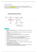
Atherosclerosis:
Progressive hardening of arteries due to the build up of plaque and thus narrowing of the
lumen
Gradual Deposition of (LDL) Cholesterol underneath the endothelium that lines the entire
vasculature
Clinically silent disease that happens over decades
Progressive - intimal thickening progresses, until later in the disease when luminal
obstruction occurs.
In timeline of atherosclerosis, the compensatory enlargement as intimal thickening
progresses, until later in the disease when luminal obstruction occurs.
If a clot forms inhibits oxygenated blood to the heart and cardiac myoctes die due to
necrosis
Cholesterol deposited underneath endothelium , leukocytes ingest cholesterol
Lesions can either grow – angina
Lesions can rupture, contents of plaque enter bloodstream , platelets recognise as foreign
causing blood clot, artery thus blocked
Leisions white colour due to calcium deposition
Precipitating thrombi that obstruct blood flow to the heart (coronary heart disease), brain
(ischemic stroke), or lower extremities (peripheral vascular disease).
,Atherogenesis involves:
1 Endothelial dysfunction, with altered NO biosynthesis which predisposes to
atherosclerosis.
2 Injury of dysfunctional endothelium, which leads to expression of adhesion molecules.
This encourages monocyte attachment and migration of monocytes from the lumen into
the intima. Lesions have a predilection for regions of disturbed flow such as the origins
of aortic branches.
3 Low-density lipoprotein (LDL) cholesterol transport into the vessel wall. Endothelial
cells and monocytes/macrophages generate free radicals that oxidise LDL (oxLDL),
resulting in lipid peroxidation.
4 oxLDL uptake by macrophages via ‘scavenger’ receptors. Such macrophages are
called foam cells because of their ‘foamy’ histological appearance, resulting from
accumulation of cytoplasmic lipid, and are characteristic of atheroma. Uptake of oxLDL
activates macrophages which release proinflammatory cytokines.
5 Subendothelial accumulation of foam cells and T lymphocytes to form fatty streaks .
6 Protective mechanisms, for example cholesterol mobilisation from the artery wall and
transport in plasma as high-density lipoprotein (HDL) cholesterol, termed ‘reverse
cholesterol transport’.
7 Cytokine and growth factor release by activated platelets, macrophages and
endothelial cells, causing proliferation of smooth muscle and deposition of connective
tissue components. This inflammatory fibroproliferative response leads to a dense
fibrous cap overlying a lipid-rich core, the whole structure comprising the atheromatous
plaque.
8 Plaque rupture , which provides a substrate for thrombosis. The presence of large
numbers of macrophages predisposes to plaque rupture, whereas vascular smooth
muscle and matrix proteins stabilise the plaque.
Thrombosis of a disrupted atheroma: weakening of the fibrous cap
,Erosion/rupture of the fibrous cap (e.g. by proteolytic enzyme release by inflammatory cells)
Most coronary syndromes are caused by thrombosis of a disrupted atheroma, which can
result from weakening of the fibrous cap and enhanced thrombogenicity of the lipid core.
The inflammatory cells can send molecular messages to the smooth muscle cells (interferon-
g) that inhibit the ability of this cell type to synthesize new collagen to strengthen the
plaque's fibrous cap.
In addition, the inflammatory cells can release proteolytic enzymes capable of degrading
collagen and other structurally important constituents of the plaque's fibrous cap.
Thus, when there is inflammation in the intima, the collagen responsible for the integrity of
the plaque's fibrous cap is under double attack, subject to both decreased synthesis and
increased degradation.
This sets the stage for plaque disruption.
The inflammatory cells also are responsible for signaling and producing increased quantities
of tissue factor, a potent procoagulant deemed responsible for thrombosis of ruptured
plaques.
Erosion or rupture of “fibrous cap” allows contact of blood with thrombogenic lipid core
Platelet aggregation and thrombus formation occludes coronary artery
Mechanisms of atherogenesis:
• Endothelial dysfunction and lipid accumulation
• The contribution of leukocytes to atherosclerosis
• Stable vs unstable plaques
NO inhibits thrombosis and maintains normal blood flow through the coronary arteries. Is an
important signaling molecule involved in vascular smooth muscle cell relaxation.
Atherosclerosis is a classic example of “sterile inflammation” - Where there are no pathogens but a
problem within our own bodies
, Atherosclerotic plaque formation involves 5 steps:
1. low density lipoprotein (LDL) accumulation in the intima;
2. oxidation of LDL;
3. recruitment of monocytes-macrophages;
4. uptake of oxidized LDL by macrophage scavenger receptors, and transformation of macrophages
into foam cells;
5. formation of a fibrous cap containing smooth muscle cells, which permits stabilization of the
plaque.
At each step of this process, inflammatory cytokines are implicated making the atherosclerotic
process a chronic inflammatory disease. (sterile inflammation)
Gradual deposition of cholesterol underneath single layer of endothelial cells
Macrophages are real culprit cells (monocytes are present in blood, differentiate into
macrophages in tissue)
Macrophages engulf oxidised LDL cholesterol – become foam cells
Foam cells aren’t removed and release inflammatory cytokines like Il1beta
Pro Il1beta is inactive form
Macrophages have inflammasome that is involved in processing Pro Il1beta into active form
– happens in macrophages that overconsume LDL
Stimulate endothelial cells that attract more monocytes to enter
Vicious cycle of foam cells attracting more macrophages
Defective egress – cells burst open, necrosis, close by immune cells signal immune cell
recruitment
Gradual growth of lesion that comes about by continued recruitment of cells, engulfment of
LDL and necrosis
Forms a necrotic core that builds up
Held during the summer examination period, this will be a 3 hour paper consisting of EIGHT
questions requiring essay style answers, from which you must choose THREE to answer. Each essay
will contribute 33.3% of the final mark for the paper.
Atherosclerosis:
Progressive hardening of arteries due to the build up of plaque and thus narrowing of the
lumen
Gradual Deposition of (LDL) Cholesterol underneath the endothelium that lines the entire
vasculature
Clinically silent disease that happens over decades
Progressive - intimal thickening progresses, until later in the disease when luminal
obstruction occurs.
In timeline of atherosclerosis, the compensatory enlargement as intimal thickening
progresses, until later in the disease when luminal obstruction occurs.
If a clot forms inhibits oxygenated blood to the heart and cardiac myoctes die due to
necrosis
Cholesterol deposited underneath endothelium , leukocytes ingest cholesterol
Lesions can either grow – angina
Lesions can rupture, contents of plaque enter bloodstream , platelets recognise as foreign
causing blood clot, artery thus blocked
Leisions white colour due to calcium deposition
Precipitating thrombi that obstruct blood flow to the heart (coronary heart disease), brain
(ischemic stroke), or lower extremities (peripheral vascular disease).
,Atherogenesis involves:
1 Endothelial dysfunction, with altered NO biosynthesis which predisposes to
atherosclerosis.
2 Injury of dysfunctional endothelium, which leads to expression of adhesion molecules.
This encourages monocyte attachment and migration of monocytes from the lumen into
the intima. Lesions have a predilection for regions of disturbed flow such as the origins
of aortic branches.
3 Low-density lipoprotein (LDL) cholesterol transport into the vessel wall. Endothelial
cells and monocytes/macrophages generate free radicals that oxidise LDL (oxLDL),
resulting in lipid peroxidation.
4 oxLDL uptake by macrophages via ‘scavenger’ receptors. Such macrophages are
called foam cells because of their ‘foamy’ histological appearance, resulting from
accumulation of cytoplasmic lipid, and are characteristic of atheroma. Uptake of oxLDL
activates macrophages which release proinflammatory cytokines.
5 Subendothelial accumulation of foam cells and T lymphocytes to form fatty streaks .
6 Protective mechanisms, for example cholesterol mobilisation from the artery wall and
transport in plasma as high-density lipoprotein (HDL) cholesterol, termed ‘reverse
cholesterol transport’.
7 Cytokine and growth factor release by activated platelets, macrophages and
endothelial cells, causing proliferation of smooth muscle and deposition of connective
tissue components. This inflammatory fibroproliferative response leads to a dense
fibrous cap overlying a lipid-rich core, the whole structure comprising the atheromatous
plaque.
8 Plaque rupture , which provides a substrate for thrombosis. The presence of large
numbers of macrophages predisposes to plaque rupture, whereas vascular smooth
muscle and matrix proteins stabilise the plaque.
Thrombosis of a disrupted atheroma: weakening of the fibrous cap
,Erosion/rupture of the fibrous cap (e.g. by proteolytic enzyme release by inflammatory cells)
Most coronary syndromes are caused by thrombosis of a disrupted atheroma, which can
result from weakening of the fibrous cap and enhanced thrombogenicity of the lipid core.
The inflammatory cells can send molecular messages to the smooth muscle cells (interferon-
g) that inhibit the ability of this cell type to synthesize new collagen to strengthen the
plaque's fibrous cap.
In addition, the inflammatory cells can release proteolytic enzymes capable of degrading
collagen and other structurally important constituents of the plaque's fibrous cap.
Thus, when there is inflammation in the intima, the collagen responsible for the integrity of
the plaque's fibrous cap is under double attack, subject to both decreased synthesis and
increased degradation.
This sets the stage for plaque disruption.
The inflammatory cells also are responsible for signaling and producing increased quantities
of tissue factor, a potent procoagulant deemed responsible for thrombosis of ruptured
plaques.
Erosion or rupture of “fibrous cap” allows contact of blood with thrombogenic lipid core
Platelet aggregation and thrombus formation occludes coronary artery
Mechanisms of atherogenesis:
• Endothelial dysfunction and lipid accumulation
• The contribution of leukocytes to atherosclerosis
• Stable vs unstable plaques
NO inhibits thrombosis and maintains normal blood flow through the coronary arteries. Is an
important signaling molecule involved in vascular smooth muscle cell relaxation.
Atherosclerosis is a classic example of “sterile inflammation” - Where there are no pathogens but a
problem within our own bodies
, Atherosclerotic plaque formation involves 5 steps:
1. low density lipoprotein (LDL) accumulation in the intima;
2. oxidation of LDL;
3. recruitment of monocytes-macrophages;
4. uptake of oxidized LDL by macrophage scavenger receptors, and transformation of macrophages
into foam cells;
5. formation of a fibrous cap containing smooth muscle cells, which permits stabilization of the
plaque.
At each step of this process, inflammatory cytokines are implicated making the atherosclerotic
process a chronic inflammatory disease. (sterile inflammation)
Gradual deposition of cholesterol underneath single layer of endothelial cells
Macrophages are real culprit cells (monocytes are present in blood, differentiate into
macrophages in tissue)
Macrophages engulf oxidised LDL cholesterol – become foam cells
Foam cells aren’t removed and release inflammatory cytokines like Il1beta
Pro Il1beta is inactive form
Macrophages have inflammasome that is involved in processing Pro Il1beta into active form
– happens in macrophages that overconsume LDL
Stimulate endothelial cells that attract more monocytes to enter
Vicious cycle of foam cells attracting more macrophages
Defective egress – cells burst open, necrosis, close by immune cells signal immune cell
recruitment
Gradual growth of lesion that comes about by continued recruitment of cells, engulfment of
LDL and necrosis
Forms a necrotic core that builds up