Cornell notes template
o Neuroscience
Neuroscience - Servingpsychology:
methods for physiological for study of relationship between brain and behaviour
techniques - An ideal method? Spatial resolution ( cellular level temporal
Overview of electrophysiological methods:
resolution: millisecond scale)
o Transcranial magnetic stimulation (TMS)
- Whole brain studied simultaneously
o EEG and event related potentials (ERPs)
- Non invasive
Magnetic resonance imaging: structural
- No such methodand functional (fMRI)
Neuropsychology based on - lesion
Matchstudies
existing method all with certain limitations, to the research
question
o Structural - Goals of structural imaging with non invasive methods:
(anatomical) - To study anatomy
MRI – as - To identify abnormalities (as in brain disease) to follow
opposed to development (childhood to old age)
functional - To show plasticity
MRI - Methods of interest to biological psychology examples:
- Computed tomography (CT) scans
- MRI scans – 1980: nobel prize 2003 5o Sir Peter Mansfield (1933-
2017), Uni of Nottingham and to Paul Lauterbur, 1929-2007)
- (CT and) structural MRI rely on contrast between tissue types
(white matter vs gray matter vs cerebrospinal fluid)
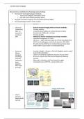
o Structural - Learning to juggle = changes to the brain, juggling requires expert
MRI for visual motion perception
biological - Clusters of statistically significant expansion of gray matter
psychology - Not to be confused with fMRI (similar display but different
(example method)
application – - Observed in volunteers who have learned to juggle
brain - Correspond to area hMT/V5, a visual motion area
plasticity
after motor
learning)
o How to
generate
structural MR
contrast
, Cornell notes template
- Functional imaging
- Goal: identify brain areas that support sensory and cognitive processes,
derive models of brain function
- Blood flow (PET/SPECT/fMRI)
- Need contrast that separated non activated vs activated tissue (in a
stimulus – or task specific way)
- Problem one: how to measure neural activity in functional contrast?
- Problem two: how to generate measurable functional contrast in
experiment?
- Problem three: how to identify functional contrast in fMRI raw data?
- Somatotopic brain activation during movement of right foot, right
elbow, right hand (fingers) and lips
- T2* contrast
o Structural MR - Depends on balance of deoxygenated to oxygenated hemoglobin (Hb)
contrast than within blood in a voxel
T1 contrast? - this is turn depends on local regulation of arterial width
o Functional
MRI (as
opposed to
structural
MRI)
o How to
measure
neural
activity in
functional
contrast:
BOLD effect
- block designs
o Neuroscience
Neuroscience - Servingpsychology:
methods for physiological for study of relationship between brain and behaviour
techniques - An ideal method? Spatial resolution ( cellular level temporal
Overview of electrophysiological methods:
resolution: millisecond scale)
o Transcranial magnetic stimulation (TMS)
- Whole brain studied simultaneously
o EEG and event related potentials (ERPs)
- Non invasive
Magnetic resonance imaging: structural
- No such methodand functional (fMRI)
Neuropsychology based on - lesion
Matchstudies
existing method all with certain limitations, to the research
question
o Structural - Goals of structural imaging with non invasive methods:
(anatomical) - To study anatomy
MRI – as - To identify abnormalities (as in brain disease) to follow
opposed to development (childhood to old age)
functional - To show plasticity
MRI - Methods of interest to biological psychology examples:
- Computed tomography (CT) scans
- MRI scans – 1980: nobel prize 2003 5o Sir Peter Mansfield (1933-
2017), Uni of Nottingham and to Paul Lauterbur, 1929-2007)
- (CT and) structural MRI rely on contrast between tissue types
(white matter vs gray matter vs cerebrospinal fluid)
o Structural - Learning to juggle = changes to the brain, juggling requires expert
MRI for visual motion perception
biological - Clusters of statistically significant expansion of gray matter
psychology - Not to be confused with fMRI (similar display but different
(example method)
application – - Observed in volunteers who have learned to juggle
brain - Correspond to area hMT/V5, a visual motion area
plasticity
after motor
learning)
o How to
generate
structural MR
contrast
, Cornell notes template
- Functional imaging
- Goal: identify brain areas that support sensory and cognitive processes,
derive models of brain function
- Blood flow (PET/SPECT/fMRI)
- Need contrast that separated non activated vs activated tissue (in a
stimulus – or task specific way)
- Problem one: how to measure neural activity in functional contrast?
- Problem two: how to generate measurable functional contrast in
experiment?
- Problem three: how to identify functional contrast in fMRI raw data?
- Somatotopic brain activation during movement of right foot, right
elbow, right hand (fingers) and lips
- T2* contrast
o Structural MR - Depends on balance of deoxygenated to oxygenated hemoglobin (Hb)
contrast than within blood in a voxel
T1 contrast? - this is turn depends on local regulation of arterial width
o Functional
MRI (as
opposed to
structural
MRI)
o How to
measure
neural
activity in
functional
contrast:
BOLD effect
- block designs