CELL STRUCTURE
(a) Use of microscopy to observe and investigate different types of cell and cell
structure in a range of eukaryotic organisms.
● To include an appreciation of the images produced by a range of microscopes,
light microscope, transmission electron microscope, scanning electron
microscope and laser scanning confocal microscope.
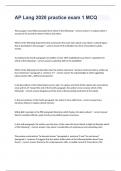
Light microscope
Light microscopes are the most ubiquitous of the all the microscopes because they
were the first-developed, they are cheap, portable, able to study whole living
specimens, and are easy to use.
The compound light microscope has two lenses- the objective lense, which is
placed near the specimen, and the eyepiece lens, through which the specimen is
viewed. The objective lens produces a magnified image, which is magnified again by
the the eyepiece lens. This objective/eyepiece lens configuration allows for much
higher magnification and reduced chromatic aberration than that in a simple light
microscope. Illumination is usually provided from underneath the sample, through
opaque sample may be illuminated from above.
Optical microscopes allow magnification of up to x1500, which enables us to
see clearly some of the larger structures inside cells. However, because their
resolution is limited, they cannot magnify any higher while still giving a clear image.
This is because they use visible light, a part of the electromagnetic spectrum, that has
a wavelength of 400-700 nm. So it has a resolving power of about 200 nm. This means
they cannot see many organelles, ribosomes, for example, have a 30 nm diameter, so
are not resolved.
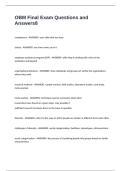
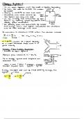
Electron Microscopes
For light microscopes, it is the wavelength of the light that becomes the limiting
factor, because the resolving power can not be reduced below 200 nm. However, in
electron microscopy, a beam of fast-travelling electrons is used, with a wavelength of
0.004nm, meaning that they have much greater resolution than optical microscopes
and can be used to give clear and highly magnified images.
The electrons are fired from a cathode and focused by magnets rather than
glass lenses, onto a screen or photographic plate.
The use of electron, however, requires preparation involving killing the cell, because a
vacuum must be used. This is not only expensive, but can give rise to artefacts,
changes in the ultrastructure as a result of the preparation. They can be seen in the
loss of continuity of membranes, distortion of organelles and empty spaces between
the cytoplasm of cells.
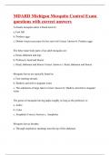
● Transmission electron microscopes:
Use a beam of electrons, transmitted through a specimen and focused to
produce an image, the same principle as light microscopy. It has the best
(a) Use of microscopy to observe and investigate different types of cell and cell
structure in a range of eukaryotic organisms.
● To include an appreciation of the images produced by a range of microscopes,
light microscope, transmission electron microscope, scanning electron
microscope and laser scanning confocal microscope.
Light microscope
Light microscopes are the most ubiquitous of the all the microscopes because they
were the first-developed, they are cheap, portable, able to study whole living
specimens, and are easy to use.
The compound light microscope has two lenses- the objective lense, which is
placed near the specimen, and the eyepiece lens, through which the specimen is
viewed. The objective lens produces a magnified image, which is magnified again by
the the eyepiece lens. This objective/eyepiece lens configuration allows for much
higher magnification and reduced chromatic aberration than that in a simple light
microscope. Illumination is usually provided from underneath the sample, through
opaque sample may be illuminated from above.
Optical microscopes allow magnification of up to x1500, which enables us to
see clearly some of the larger structures inside cells. However, because their
resolution is limited, they cannot magnify any higher while still giving a clear image.
This is because they use visible light, a part of the electromagnetic spectrum, that has
a wavelength of 400-700 nm. So it has a resolving power of about 200 nm. This means
they cannot see many organelles, ribosomes, for example, have a 30 nm diameter, so
are not resolved.
Electron Microscopes
For light microscopes, it is the wavelength of the light that becomes the limiting
factor, because the resolving power can not be reduced below 200 nm. However, in
electron microscopy, a beam of fast-travelling electrons is used, with a wavelength of
0.004nm, meaning that they have much greater resolution than optical microscopes
and can be used to give clear and highly magnified images.
The electrons are fired from a cathode and focused by magnets rather than
glass lenses, onto a screen or photographic plate.
The use of electron, however, requires preparation involving killing the cell, because a
vacuum must be used. This is not only expensive, but can give rise to artefacts,
changes in the ultrastructure as a result of the preparation. They can be seen in the
loss of continuity of membranes, distortion of organelles and empty spaces between
the cytoplasm of cells.
● Transmission electron microscopes:
Use a beam of electrons, transmitted through a specimen and focused to
produce an image, the same principle as light microscopy. It has the best