Biomedical Imaging – Microscopy
Introduction
1. The human eye is a powerful detector
The eye has its own imaging microscope
• Very large dynamic range
- adaptive: viewing in dark vs. light
- Because of a variable pupil size that is controlled by an aperture (iris) that can close as
more light comes in
- It has hundreds of millions of pixels
• Retina (= light-sensitive plate): Very large screen with individual light-sensitive pigment cells/
photoreceptors (cones and rods)
- photoreceptors: about 10 million
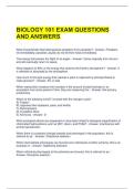
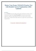
Anatomy of the human eye
• biological lens that can change = tuneable lens using a muscle
• lens, which is covered by an opening (pupil) that is also tuneable
- light passes through the pupil (light reaches the sensitive plate all the way at the back
= retina)
• an diaphragm that can open when it is darker and close when it is lighter, which is of course
the iris
The human eye has limited resolving power
➔ EXAM: what is resolution? = The ability to distinguish objects = about 0.1 mm (this is quite
limited)
We are unable to distinguish objects closer than 0.1 mm apart. You can still see something, but
it looks blurry
Resolution can be boosted by the use of lenses
• A lens makes something bigger so that it is still distinguishable to humans
• A lens magnifies objects and therefore is going to increase the distance between objects
- Problem of 1 lens: image is on the wrong side
• Concept of modern microscope → using 2 lenses = compound microscope = inverted telescope
• !! A microscope is nothing but a combination of at least 2 lenses (nowadays many more), which
actually makes magnification possible and therefore realizes a large distinctive power
• revolution in microscopy comes along with the revolution in cell biology (otherwise, we would
never have known that there was such a thing as a cell, nor had any knowledge of its
substructures) → important fact to know
,Basics of light microscopy (1)
A microscope is an instrument designed to make fine details visible. The microscope must accomplish
three tasks:
• Produce a magnified image of the specimen (magnification)
• Separate the details in the image (resolution)
• Render the details visible to the eye, camera or other imaging device (contrast)
1. The wave-like properties of light
Light is a wave, or at least can be considered a wave → thus has different properties which are
consistent with the wave theory
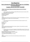
Visible light is a part of the electromagnetic spectrum
• When we talk about waves, we are talking about electromagnetic radiation. Electromagnetic
radiation waves are fluctuations of electric and magnetic fields, but it can also be described in
terms of a stream of photons, which are massless particles each traveling with wavelike
properties at the speed of light.
• the difference of radiation is in the wavelength: radio waves have a very long wavelength →
cosmic waves have a very short wavelength (the shorter the wavelength, the more energy)
• visible light (In light microscopy): 400 nm - 700 nm
Blue - green - yellow – red
- before this (< 400 nm) : UV
- after this (> 700 nm): IR
• Every electromagnetic wave is a sinus with an amplitude, phase, frequency ...
Visible light slows down in media other than vacuum
• A wave has a certain speed in a certain medium
• In vacuum the wave goes fastest because it is not stopped by anything
- Denser medium (denser relative to vacuum) → wave slows down (in air, water, oil)
- Anything denser than air (n = 1) will have a higher refractive index
➢ Water: n = 1,3
➢ Oil: n = 1,5
• From the moment the refractive index increases, light slows down
→ Refractive index n = speed of light in vacuum / speed in medium
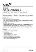
Refraction: light bends at an interface
• index of refraction increases = refraction toward the normal
→ When light encounters a medium with a higher index of refraction, light starts to deflect
toward the normal, so the angle decreases
- normal = the line perpendicular to the surface
• Snell's Law: the ratio of the refractive index' is inversely proportional to the ratio of the sine of
the angles
,Refraction depends on the wavelength = dispersion
• Refraction is wavelength-specific (color-dependent or wavelength-dependent) →so each color
refracts a little differently
- shortest wavelengths refract the hardest (UV)
- longest wavelengths break less hard (IR)
- → this is also the reason why on a fall night you see red light the longest, the UV and
all other colors are already broken down
• = Dispersion (= spreading out the different wavelengths)
Above a critical angle, light reflects
• Full reflection may also occur → angle at which light strikes = angle at which light is reflected
- angle of incidence = angle of reflection
• Not every medium will reflect perfectly (depends on consistency, density, etc.).
- A very dense medium, e.g. a metal, will reflect very strongly
Coherent light spreads out when passing a narrow slit
• bending: when a wave encounters an object or a small opening, the wave will deflect
= diffraction (you also notice this when you throw a stone into the water)
• opening = large, wave will be able to continue the same way (approximately) → small
opening: wave will deflect harder
• So: As long as the aperture is large enough, that wave keeps going nice and straight and we get
darkening on the outside
Opening too small (smaller than wavelength of the light) = deflection where you get most of
the light in the centre and a diffraction pattern in certain places
Diffraction generates a characteristic pattern
• You have 1 big peak and then min, max, min, ect. → diffraction pattern
• This is partly because light will not arrive everywhere at the same time, and on the other hand
because the waves start interacting with each other because they are bended (= interference)
→ Diffraction is a specific case of interference
Interference happens when waves meet
Interference converts phase into amplitude changes
, • Constructive interference: 2 waves are in phase and will therefore amplify each other
= 1 wave with exactly the same wavelength but double the amplitude
• Destructive interference: phase shift of exactly the same wave by 180 degrees (= about half a
wave)
→ maxima and minima of the two waves will overlap and cancel each other out (= 'flat line')
2. Lens theory
• theoretical basis: a perfectly convex lens (= doesn't exist)
• every lens is going to be able to focus light → place where the light is focused = focal point
• The distance at which that point lies = the focal distance
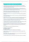
• rules of light rays
- Rule 1: Parallel rays converge at the focal point
- Rule 2: Rays from the focal plane exit in parallel
- Rule 3: Rays that pass through the centre are unaffected
Calculating the magnification
• d0 = the distance of the object from the lens, di = distance of the image from the lens, f = focal
distance
+ see exercises slides 17 – 19
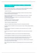
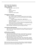
!! special case - Calculating the magnification for d0 = f
• When d0 = f, image is at infinity: you get no image here, object
is not focused anywhere → image is at infinity. There is an
image, but we cannot grasp it (virtual, intangible)
• We can still make this appear by using a second lens. That lens
can then focus the parallel beams to form an image
• This is often used in microscopy to create space. As long as the
image travels in parallel beams, we can actually create
distance and slide all sorts of things in between = typical
property of infinity optics (i.e. this gives us more distance)
Introduction
1. The human eye is a powerful detector
The eye has its own imaging microscope
• Very large dynamic range
- adaptive: viewing in dark vs. light
- Because of a variable pupil size that is controlled by an aperture (iris) that can close as
more light comes in
- It has hundreds of millions of pixels
• Retina (= light-sensitive plate): Very large screen with individual light-sensitive pigment cells/
photoreceptors (cones and rods)
- photoreceptors: about 10 million
Anatomy of the human eye
• biological lens that can change = tuneable lens using a muscle
• lens, which is covered by an opening (pupil) that is also tuneable
- light passes through the pupil (light reaches the sensitive plate all the way at the back
= retina)
• an diaphragm that can open when it is darker and close when it is lighter, which is of course
the iris
The human eye has limited resolving power
➔ EXAM: what is resolution? = The ability to distinguish objects = about 0.1 mm (this is quite
limited)
We are unable to distinguish objects closer than 0.1 mm apart. You can still see something, but
it looks blurry
Resolution can be boosted by the use of lenses
• A lens makes something bigger so that it is still distinguishable to humans
• A lens magnifies objects and therefore is going to increase the distance between objects
- Problem of 1 lens: image is on the wrong side
• Concept of modern microscope → using 2 lenses = compound microscope = inverted telescope
• !! A microscope is nothing but a combination of at least 2 lenses (nowadays many more), which
actually makes magnification possible and therefore realizes a large distinctive power
• revolution in microscopy comes along with the revolution in cell biology (otherwise, we would
never have known that there was such a thing as a cell, nor had any knowledge of its
substructures) → important fact to know
,Basics of light microscopy (1)
A microscope is an instrument designed to make fine details visible. The microscope must accomplish
three tasks:
• Produce a magnified image of the specimen (magnification)
• Separate the details in the image (resolution)
• Render the details visible to the eye, camera or other imaging device (contrast)
1. The wave-like properties of light
Light is a wave, or at least can be considered a wave → thus has different properties which are
consistent with the wave theory
Visible light is a part of the electromagnetic spectrum
• When we talk about waves, we are talking about electromagnetic radiation. Electromagnetic
radiation waves are fluctuations of electric and magnetic fields, but it can also be described in
terms of a stream of photons, which are massless particles each traveling with wavelike
properties at the speed of light.
• the difference of radiation is in the wavelength: radio waves have a very long wavelength →
cosmic waves have a very short wavelength (the shorter the wavelength, the more energy)
• visible light (In light microscopy): 400 nm - 700 nm
Blue - green - yellow – red
- before this (< 400 nm) : UV
- after this (> 700 nm): IR
• Every electromagnetic wave is a sinus with an amplitude, phase, frequency ...
Visible light slows down in media other than vacuum
• A wave has a certain speed in a certain medium
• In vacuum the wave goes fastest because it is not stopped by anything
- Denser medium (denser relative to vacuum) → wave slows down (in air, water, oil)
- Anything denser than air (n = 1) will have a higher refractive index
➢ Water: n = 1,3
➢ Oil: n = 1,5
• From the moment the refractive index increases, light slows down
→ Refractive index n = speed of light in vacuum / speed in medium
Refraction: light bends at an interface
• index of refraction increases = refraction toward the normal
→ When light encounters a medium with a higher index of refraction, light starts to deflect
toward the normal, so the angle decreases
- normal = the line perpendicular to the surface
• Snell's Law: the ratio of the refractive index' is inversely proportional to the ratio of the sine of
the angles
,Refraction depends on the wavelength = dispersion
• Refraction is wavelength-specific (color-dependent or wavelength-dependent) →so each color
refracts a little differently
- shortest wavelengths refract the hardest (UV)
- longest wavelengths break less hard (IR)
- → this is also the reason why on a fall night you see red light the longest, the UV and
all other colors are already broken down
• = Dispersion (= spreading out the different wavelengths)
Above a critical angle, light reflects
• Full reflection may also occur → angle at which light strikes = angle at which light is reflected
- angle of incidence = angle of reflection
• Not every medium will reflect perfectly (depends on consistency, density, etc.).
- A very dense medium, e.g. a metal, will reflect very strongly
Coherent light spreads out when passing a narrow slit
• bending: when a wave encounters an object or a small opening, the wave will deflect
= diffraction (you also notice this when you throw a stone into the water)
• opening = large, wave will be able to continue the same way (approximately) → small
opening: wave will deflect harder
• So: As long as the aperture is large enough, that wave keeps going nice and straight and we get
darkening on the outside
Opening too small (smaller than wavelength of the light) = deflection where you get most of
the light in the centre and a diffraction pattern in certain places
Diffraction generates a characteristic pattern
• You have 1 big peak and then min, max, min, ect. → diffraction pattern
• This is partly because light will not arrive everywhere at the same time, and on the other hand
because the waves start interacting with each other because they are bended (= interference)
→ Diffraction is a specific case of interference
Interference happens when waves meet
Interference converts phase into amplitude changes
, • Constructive interference: 2 waves are in phase and will therefore amplify each other
= 1 wave with exactly the same wavelength but double the amplitude
• Destructive interference: phase shift of exactly the same wave by 180 degrees (= about half a
wave)
→ maxima and minima of the two waves will overlap and cancel each other out (= 'flat line')
2. Lens theory
• theoretical basis: a perfectly convex lens (= doesn't exist)
• every lens is going to be able to focus light → place where the light is focused = focal point
• The distance at which that point lies = the focal distance
• rules of light rays
- Rule 1: Parallel rays converge at the focal point
- Rule 2: Rays from the focal plane exit in parallel
- Rule 3: Rays that pass through the centre are unaffected
Calculating the magnification
• d0 = the distance of the object from the lens, di = distance of the image from the lens, f = focal
distance
+ see exercises slides 17 – 19
!! special case - Calculating the magnification for d0 = f
• When d0 = f, image is at infinity: you get no image here, object
is not focused anywhere → image is at infinity. There is an
image, but we cannot grasp it (virtual, intangible)
• We can still make this appear by using a second lens. That lens
can then focus the parallel beams to form an image
• This is often used in microscopy to create space. As long as the
image travels in parallel beams, we can actually create
distance and slide all sorts of things in between = typical
property of infinity optics (i.e. this gives us more distance)