NCLE Complete Review Questions & Answers Solved 100%
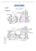
Adnexa Oculi - Answer are the accessory structure that are considered part of the eye, but not part of the eyeball itself. these structures include: eyelids, eyelashes, tears, lacrimal apparatus, accessory glands and extrinsic muscles and orbit. everything outside of the eyeball. Meibomian Glands - Answer Glands of the eyelids. sebaceous gland in the eyelid behind the gray line Glands of Zeiss - Answer Glands of the eyelids. sebaceous oily gland and is located around the eyelid cilia and follicles of the eyelashes Glands of Moll - Answer Glands of the eyelids. are sweat glands Glands of Wolfring and Krause - Answer Glands of the eyelids. located inside the lids near the fornix and are accessory glands of the lacrimal gland Lacrimal gland - Answer Glands of the eyelids. secretes tears Goblet Cells - Answer Glands of the eyelids. secrete mucin and are found in the lid conjunctiva Conjunctiva - Answer Lines the inside of the eyelids and covers the sclera Has 3 parts: Palpebral, Bulbar & Fornix palpebral conjunctiva - Answer covers inner surface of the eyelids Bulbar Conjunctiva - Answer covers the anterior part of the sclera. Helps lubricate the eye by producing tears and mucus Tear Film Functions - Answer 3 Layers (Sebaceous/Lipid, Aqueous/Lacrimal, Mucoid/Mucin) Lipid - Answer fatty Aqueous - Answer nutrients Mucoid - Answer wetting and lubrication Break-up time - Answer (BUT) - Interval between a complete blink and the first randomly distributed dry spot (10-40 seconds in normal). under 10 seconds could indicate marginal dry eye. fluorescein is used for this test Schirmer Test - Answer Determines if eye produces enough tears via placing a strip in the lower eyelid. 35-40 mm of wetting from the lid indicates normal lacrimal secretion inserted in eye 5mm strip Rose Bengal - Answer Reddish-purple dye used to detect damaged superficial corneal and conjunctival cells Jones Test - Answer Evaluates tear drainage system function. This test measures the length of time for fluorescein dye to appear inside the nose after being instilled in the cornea. Upper lashes - Answer are longer and curl up and there about 100 - 150 lashes Bottom lashes - Answer turn downward and number about 50 - 75 Corneal Topography - Answer Transparent and Avascular- (no blood). Main Refracting Surface of the eye. 75% of refraction takes place at the cornea. Convex, aspheric and smooth and tends to flatten toward the periphery. Average Refractive Power Of The Cornea - Answer is about +43.00 D *** Average Corneal Thickness - Answer is about .52mm. .53 in center, thinner and more rounded. .71 thicker and smaller at the periphery Horizontal Visible Iris Diameter - Answer 11.5 mm x 10.5 mm. HVID- normal size of the iris fitting for the contact to fit this area Apical Zone - Answer Is a Zone of the Cornea. Regular in shape Largest in center. Average size varies about 4-6mm. Referred to as Corneal Cap. Displaced up and in nasally. Apical Zone Points - Answer Visual Center, Apex & Geometric Center of the Cornea. Visual Center - Answer the point where the visual axis passes through. Part of the Apical zone. Apex - Answer steepest point on the cornea usually displaced up and in from the geometric center. Part of the Apical zone. Geometric Center Of The Cornea - Answer Part of the Apical zone. The intersection f widest ad shortest diameter of the cornea Transition Zone - Answer Is a Zone of the Cornea. Area between the Apical zone and the limbus. Area of the cornea with the greatest aspheric curvature. In this zone the cornea flattens out more temporally than nasally. 1 mm adjacent to the sclera Limbal Zone - Answer Is a Zone of the Cornea. Area near the periphery of the cornea about 1 mm adjacent to the sclera. Area is not well defined and blends into the Transition Zone of the cornea Sulcus - Answer Depression or "ditch" around the cornea and is divides the cornea from the sclera. Usually referred to as the external boundary of the cornea Layers of the Cornea - Answer Epithelium, Bowman's Membrane, stroma, Descemet's Membrane, Endothelium Epithelium - Answer Outermost layer of the cornea and is 5-6 layers thick (has 3 cell layers). Will repair itself. 3 cell layers: Squamous (flat), Wing cell, Basal cell layer (inner most layer). Bowman's Membrane - Answer Thin elastic acellular membrane of stromal collagen if damaged will scar. Cannot be separated from the stroma. Stroma - Answer middle layer of the cornea and makes up about 90% of total corneal thickness. If damaged, the stroma will leave a scar. Descemet's Membrane - Answer Elastic membrane secreted by the endothelium. Will reform if damaged. Can rupture and edges curl up under slit lamp observation. Endothelium - Answer innermost layer of the cornea. Cells are "hexagonal in shape. Cells do not regenerate when damaged. Single layer of cells for maintaining the integrity of the corneal from within. Looks like an endothelial mosaic. Polymegethsim - Answer variation in cell size, Endothelium Disorders Polymorphism - Answer variation in cell shape, Endothelium Disorders Endothelial Guttata - Answer deposits on the endothelium indicating endothelial dysfunction, Endothelium Disorders Corneal Transparency and Metabolism - Answer if there is blockage it can lead to swelling. do not want to put on an eye that can lead to edema. Osmosis, Endothelial or Metabolic Pump. Corneal Deturgescence - Answer State of dehydration, 25% 30% dehydration. Glucose - Answer form of energy is supplied to the endothelium by the aqueous humor ATP - Answer (Adenosine triphosphate) is formed when glucose breaks down into the nucleus of the cells creating a pumping action, therefore maintains the proper water balance in the cornea. the most active layers for the metabolism are. sources of nutrients, glucose, oxygen, amino acids. corneal hydration. the state of relative dehydration that is necessary for corneal transparency. normal water content is a constant 80%. Edema - Answer Swelling of corneal tissue and results when forces normally dehydrating the cornea are overcome by forces driving water into the cornea. Instrumentation to look at an Edema - Answer Pachometer, Keratometry, Slit Lamp Gross Edema - Answer reversible form of edema, swelling of epithelial cells Microcystic Edema - Answer Non-reversible edema that involves cell death at the epithelium. Caused by OWS (over wearing syndrome) painful, causes photophobia Anterior Chamber - Answer Posterior to cornea and anterior to iris, Contains aqueous humor, Trabecular Meshwork- Provides an exit for the aqueous humor, Canal of Schlemm- After aqueous flows through the trabecular meshwork, excess aqueous flows to the Canal of Schlemm. Remembering Anterior vs Posterior - Answer Anterior Chamber- A front of the alphabet it is in front. Posterior chamber- end, closer to the end of the alphabet. Posterior Chamber - Answer Behind the iris and bounded by the posterior iris surface the lens the anterior vitreous and the ciliary body Aqueous Humor - Answer Provides oxygen and glucose to cornea and lens Sclera - Answer (Fibrous Tunic) - Outermost tunic made up of fibrous connective tissue-Known as the white of the eye The Iris - Answer Filters light and UV rays due to pigmentation. Controls light by dilating and constricting pupil Pupil size - Answer Varies with individual (average adult 3-4 mm.). Changes size with intensity of ricts during sleep to eliminate light stimulus. constricts during accommodation palpebral fissure - Answer the elliptical open space between the eyelids Choroid - Answer posterior portion of the Uveal tract. Composed of blood vessels and lies between the sclera and retina. It provides blood supply for the outer cells of the retina. Crystalline Lens - Answer located immediately behind the iris. Clear, membrane-like structure that is quite elastic- holds ligaments in place Ciliary Muscle - Answer band like structure that encircles the inside of the eye from the iris root to the anterior edge of the retina attached to the ora serrata (accommodation process) Retina - Answer innermost layer of the eye, visual pathway to the brain, converts light entering the eye via nerve impulses, we see with our brain the eyes collect the information to do so. Photoreceptors - Answer Rods & Cones Rod - Answer Type of Photoreceptor- night vision, no color vision found in periphery Cones - Answer Type of Photoreceptor- day vision, color vision (ice cream during the day with rainbow sprinkles) Ocular Pathology - Answer eye care professional must be aware of medications, diseases and viruses that can affect the fitting of contact lens. Considerations: Alcohol, Diabetes, Arthritis, HIV, General medications that may cause dry eye Hordeolum - Answer stye, blocked zeiss gland, staph infection, tender to the touch, self-resolving (warm compress). Chalazion - Answer blocked meibomian gland, painless, excision (removal) may be necessary. Blepharitis - Answer inflammation of eyelid margins, eyelid scrubs. Warm compresses, staph infection/antibiotic ointment.
Escuela, estudio y materia
- Institución
- NCLE
- Grado
- NCLE
Información del documento
- Subido en
- 30 de octubre de 2023
- Número de páginas
- 36
- Escrito en
- 2023/2024
- Tipo
- Examen
- Contiene
- Preguntas y respuestas
Temas
-
ncle
-
ncle complete review questions answers solved 10
-
adnexa oculi are the accessory structure that are
-
meibomian glands glands of the eyelids sebaceous
Documento también disponible en un lote