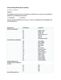
Samenvatting cardiovascular physiology
concepts
Chapter 1, introduction to cardiovascular system
Cells need metabolic substrates (e.g. O2, amino acids, glucose) and mechanism that removes
byproducts of metabolism (e.g. CO2, lactic acid).
- Single cell organisms exchange with environment through diffusion + cellular transport
system
- Most cells of large organisms limited/no exchange capacity (not in contact with
environment). Thus blood vessels cells-blood and blood-environment.
Exchange with outside environment:
- Lungs
- Gastrointestinal tract
- Kidneys
- Skin
Blood passing through lungs O2 and CO2
Blood passing intestine glucose, amino acids, fatty acids and other ingested substances to liver
additional metabolic processing.
Proper balance of water and electrolytes (e.g. Na+, K+ and Ca2+) to function. Kidneys eliminate
excessive water/electrolytes in urine.
Skin exchange of water/electrolytes (sweating), exchange of heat (byproduct of cellular
metabolism blood flow through skin regulates heat loss).
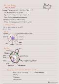
Cardiovascular system:
- Heart
- Blood vessels
- Lymphatic system exchange function in conjunction with blood vessels
Pulmonary circulation = blood flow within lungs exchange of gases between blood and alveoli.
Systemic circulation = all blood vessels within/outside of organs (excluding lungs)
Right atrium venous blood from systemic circulation. Right ventricle pulmonary circulation.
Left atrium O2 rich blood from pulmonary veins from lungs. Left ventricle ejects blood into
aorta.
Some of the fluid, along with electrolytes and small amounts of protein, leave the circulation and
enter the tissue interstitium (process termed fluid filtration). Lymphatic vessels collect excess fluid
from within the tissue interstitium and transport it back to venous circulation through lymphatic
ducts, emptying into large veins (subclavian veins above RA).
Heart side are in series with each other all blood that is pumped from RV pulmonary
circulation LA/LV systemic circulation.
Circulation of most major organ systems are in parallel. Except liver (hepatic portal system). Parallel
arrangement of major vascular beds prevents blood flow changes in 1 organ from significantly
affecting blood flow in other organs.
,Organ blood flow is not driven by the output of the heart per se, but rather by pressure generated
within the arterial system as the heart pumps blood into the vasculature (= resistance network).
(𝑎𝑟𝑡𝑒𝑟𝑖𝑎𝑙 𝑝𝑟𝑒𝑠𝑠𝑢𝑟𝑒−𝑣𝑒𝑛𝑜𝑢𝑠 𝑝𝑟𝑒𝑠𝑠𝑢𝑟𝑒)
Organ blood flow is determined by .
𝑣𝑎𝑠𝑐𝑢𝑙𝑎𝑟 𝑟𝑒𝑖𝑠𝑡𝑎𝑛𝑐𝑒 𝑜𝑓 𝑜𝑟𝑔𝑎𝑛
Pressure of CVS = mm of mercury (mm Hg) above atmospheric pressure. 1 mm Hg = 1.36 cm H2O
hydrostatic pressure).
Vascular resistance is determined by:
- Size of blood vessels
- Anatomical arrangement of vascular network
- Viscosity of blood within vasculature
RA receives blood at low pressure (venous return +/- 0 mm Hg). RA contraction into RV RV
contraction into pulmonary artery maximal pressure (systolic pressure) from 20 – 30 mm Hg. BP
falls to 10 mm Hg in pulmonary circulation. LA receives pulmonary venous blood passively flow
into LV. LV ejects blood into systemic arterial system (100 – 140 mmHg = maximal systolic pressure)
Cardiac output = amount of blood ejected with each contraction (i.e. stroke volume * heart rate).
HR is determined by specialized cells within the heart (electrical pacemakers under control of
autonomic nerves and hormones).
Besides pumping blood, the heart synthesizes several hormones atrial natriuretic peptide
important role in regulation of blood volume and blood pressure. Sensor nerve receptors associated
with heart release of antidiuretic hormone from posterior pituitary (regulates H2O loss by
kidneys).
Blood vessels constrict/dilate to:
- Regulate arterial BP
- Alter blood flow within organs
- Regulate capillary BP
- Distribute blood volume within the body.
Changes in vascular diameters by activation of vascular smooth muscle by
autonomic nerves, metabolic and biochemical signals from outside of
blood vessels, and vasoactive substances released by endothelial cells
that line blood vessels. Endothelium also produces substances that
modulate hemostasis (blood clotting) and inflammatory responses.
Organ function is dependent on circulation of blood and cardiovascular
function is dependent on function of organs. E.g. renal dysfunction
increase in blood volume hypertension or exacerbate heart failure.
Baroreceptors provide central nervous system with info regarding the
status of blood pressure in the body (through afferent neural connections
to brain). Decrease in arterial pressure stimulates heart to increase CO and constricts blood
vessels. Rapid changes in autonomic nerve activity (through sympathetic nerves) cardiovascular
adjustments.
Negative feedback process in which a deviation from some condition (e.g. normal arterial
pressure) responses that diminish deviation.
In addition to autonomic nerve activity, release of hormones help to restore arterial pressure by
action on the heart and blood vessels (increase arterial pressure by increasing blood volume through
actions on renal function). This mechanism require hours – days. Including secretion of
,catecholamines (epinephrine) by adrenal glands, renin by kidneys ( formation of angiotensin2 and
aldosteron) and release of antidiuretic hormone (vasopressin) by posterior pituitary.
Chapter 2, electrical activity of the heart
Primary function of cardiac myocytes is to contract. Electrical changes within myocytes initiate this
contraction.
Cardiac cells resting membrane potential +/- -90 mV (outside of cell is considered 0 mV). Resting
membrane potential (Em) = determined by concentrations of positively/negatively charged ions
across cell membrane, relative permeability of cell membrane to these ions, and ionic pumps that
transport ions across cell membrane.
- K+ high inside, low outside
- Na+/Ca2+ low inside, high outside
Chemical gradient (concentration difference) for K+ to diffuse out of cell, and Na+/Ca2+ into the cell.
The concentration difference is determined by activity of energy-dependent ionic pumps and
presence of impermeable, negatively charged proteins within the cell.
Equilibrium potential for K+ = Ek Nernst potential (37 degrees Celsius):
[𝐾+]𝑖
𝐸𝑘 = −61𝑙𝑜𝑔 [𝐾+}]𝑜 = -96 mV.
[K+]inside = 150 mM and [K+]outside = 4 mM.
Equilibrium potential = potential difference across membrane required to maintain the concentration
gradient across the membrane. At -96 mV: optimal K+ diffusion. The Em for ventricular myocyte is
about -90 mV net electrochemical force (= net driving force) acting on K+. Net electrochemical
driving force = +6 mV (for K+).
Equilibrium potential for Na+ = ENa:
[𝑁𝑎+]𝑖
𝐸 𝑛𝑎 = −61𝑙𝑜𝑔 [𝑁𝑎+}]𝑜 = 52 mV.
[Na+]inside = 20 mM, [Na+]outside = 145 mM..
Net electrochemical force on Na+ has 2 components:
- Na+ concentration gradient is driving sodium into cell
- Interior of resting cell is very negative large electrical force is trying to pull sodium into
cell. (-90 - +52 = -142). At rest, the permeability for Na+ is very low.
ECa2+ = +134 mV same electrochemical force as Na+.
Em is very permeable for K+ and less permeable for Na+/Ca2+ Na+/Ca2+ have little contribution
to Em.
𝑖𝑜𝑛 𝑐𝑢𝑟𝑟𝑒𝑛𝑡
Conductance (g) =𝑛𝑒𝑡 𝑒𝑙𝑒𝑐𝑡𝑟𝑜𝑐ℎ𝑒𝑚𝑖𝑐𝑎𝑙 𝑓𝑜𝑟𝑐𝑒.
Changes in Em primarily result from changes in ionic conductances.
Maintenance of ionic concentration gradients require energy (ATP hydrolysis) coupled with ionic
pumps. Whenever action potential is generated additional Na+ enters cell and additional K+
leaves. Many APs can lead to a significant change in extracellular/intracellular concentration of
K+/Na+. To maintain concentration gradients, Na+/K+ ATPase (= ATP-dependent pump) pumps Na+
out and K+ into the cell. If this pump stops working (e.g. ATP is lost under hypoxic conditions), or if
activity of pump is inhibited (cardiac glycosides, e.g. digoxin) Na+ accumulates within cell and
intracellular K+ falls more depolarized resting membrane potential. Na+/K+ ATPase pump is
electrogenic extrudes 3 Na+ for every 2 K+ entering the cell (keeps negative potential in cell).
,Inhibition of this pump causes depolarization.
In addition, increases in intracellular
Na+/extracellular K+ stimulates activity of
electrogenic Na+/K+ ATPase pump and
produce hyperpolarizing currents.
During AP, also Ca2+ enters the cell. 2
primary mechanisms to remove Ca2+ from
cells:
- ATP-dependent Ca2+ pump
actively pumps Ca2+ out of cell and
generates small negative electrogenic
potential.
- Sodium-calcium exchanger Na+
and Ca2+ are transported into
opposite directions. Can operate in
either direction across sarcolemma,
depending on Em.
o Resting cell negative Em Na+ enter cell in exchange for Ca2+ which leaves the
cell
3 Na+ for each Ca2+.
Ions move across the sarcolemma through specialized ion channels in phospholipid bilayer of cell
membrane (made of large polypeptide chains). Conformational changes in ion channel proteins
permitting ions to transverse the membrane channel/blocking ion movement.
2 general types of ion channels:
- Voltage-gated channels
open/close in response to
changes in membrane potential.
- Receptor-gated channels
open/close in response to
chemical signals operating
through membrane receptors.
E.g. ACh (NT released by vagus
nerves innervating the heart,
binds to sarcolemmal receptor
that leads to opening of special
types of potassiom channels).
Fast sodium channels (voltage):
- M gate (activation gate) is
closed
- H-gate (inactivation gate) is
open
AP activates M gate Na+, driven
by electrochemical gradient, into cell. As
M-gate open, h-gates close. M-gates
open more rapidly than h-gates close.
Closing of h-gates limits the length of
time that Na+ can enter cell. This inactivated, closed state persists throughout the repolarization
phase. Near the end of repolarization, negative membrane potential causes m-gates to close and h-
gates to open (back to initial resting, closed state).
,Activation/inactivation gates occurs when resting membrane potential is normal (-90 mV) and a rapid
depolarization occurs. How more depolarized myocyte cell is, the greater inactivated sodium
channels closing h-gates. At membrane potential -55 mV, all sodium channels are inactivated.
A cardiac cell has many sodium channels. The amount of sodium that passes when depolarization
occurs depends upon the number of sodium channel, duration of opening state and electrochemical
gradient. Each channel has slightly different voltage activation threshold and duration of open,
activated state.
Action potentials occur when membrane potential depolarizes and repolarizes back to resting state.
2 general types of cardiac APs:
- Nonpacemaker triggered by depolarizing currents from adjacent cells
- Pacemaker capable to generate spontaneous APs.
, Both types of APs differ from skeletal muscle APs. Major difference: duration of APs in a typical
nerve, the AP is +/- 1 – 2 milliseconds, in skeletal muscle cells the AP is +/- 2 – 4 milliseconds.
Duration of ventricular APs range from 200 – 400 milliseconds.
‘Fast response’ nonpacemaker APs (in atrial and ventricular myocytes, and
Purkinje fibers). 5 phases:
- Phase 4 = true resting membrane potential near equilibrium
potential for K+.
- Phase 0 = rapid depolarization when cells are depolarized from -90
mV to -70 mV (threshold voltage). It’s initiated by transient increase
in conductance of voltage-gated, fast Na+ channels.
- Phase 1 = initial repolarization caused by opening of special
type of K+ channels (transient outward) and inactivation of Na+
channels. However, because of the large increase in slow inward
gCa2+ the repolarization is delayed and AP reaches phase 2.
- Phase 2 = plateau phase inward Ca2+ movement due to long-
lasting (L-type) Ca2+ channels (open when membrane potential
depolarize to -40 mV). These cells are blocked by classical L-type
calcium channel blockers (e.g. verapamil and diliazem).
- Phase 3 = repolarization gK+ increases through delayed rectifier
K+ channels and gCA2+ decreases
Changes in Na+, Ca2+ and K+ conductances primarily determine the AP in
nonpacemaker cells.
During phase 0, 1, 2 and part of 3 the cell is refractory to initiation of new
APs = effective/absolute refractory period (ERP/ARP). In these phases h-
gates are still closed no APs. ERP act as protective mechanism in the heart by limiting frequency of
APs (therefore contractions) enables the heart have adequate time to fill/eject blood and it
prevents development of sustained, tetanic contractions (like in skeletal muscle). At the end of ERP
relative refractory period. Early in this period, suprathreshold depolarization stimuli are required
to elicit APs. Not all sodium channels have recovered, APs have a decreased phase 0 slope and a
lower amplitude.
Pacemaker cells have no true resting potentials, but generate regular, spontaneous APs. The
depolarizing current of AP is carried primarily by relatively slow, inward Ca2+ currents (L-type Ca2+
channels) instead by fast Na+ currents. The rate of depolarization is slow compared to the ‘fast
response’ nonpacemaker cells (so pacemaker AP are also called ‘slow response’). S cells in sinoatrial
(SA) node (in posterior wall of RA) primary pacemaker site. Other pacemaker cells exist within AV
node and ventricular conduction system, but firing rates are driven by higher rate of SA node
(intrinsic pacemaker activity of secondary pacemakers is suppressed by mechanisms: overdrive
suppression). The mechanism causes 2nd pacemaker to become hyperpolarized when driven at a rate
above intrinsic rate. Hyperpolarization occurs because increased AP frequency stimulates activity of
electrogenic Na+/K+-ATPase pump enhanced entry of sodium. If SA node becomes depressed
AP fail to reach 2nd pacemakers overdrive suppression ceases 2nd site takes over as pacemaker,
when this occurs the new pacemaker outside of SA node is called ectopic focus.
SA nodal AP 3 phases:
- Phase 0 = upstroke of AP depolarization primarily due to increased gCA2+ through L-type
calcium channels (at -40 mV). Rate of depolarization is slow because movement of Ca2+ is
not rapid. As membrane potential moves toward Ca2+ equilibrium transient decrease in
gK+ voltage-operated, delayed rectifier potassium channels open and increased gK+
repolarizes cell toward equilibrium potential for K+ (phase 3).
concepts
Chapter 1, introduction to cardiovascular system
Cells need metabolic substrates (e.g. O2, amino acids, glucose) and mechanism that removes
byproducts of metabolism (e.g. CO2, lactic acid).
- Single cell organisms exchange with environment through diffusion + cellular transport
system
- Most cells of large organisms limited/no exchange capacity (not in contact with
environment). Thus blood vessels cells-blood and blood-environment.
Exchange with outside environment:
- Lungs
- Gastrointestinal tract
- Kidneys
- Skin
Blood passing through lungs O2 and CO2
Blood passing intestine glucose, amino acids, fatty acids and other ingested substances to liver
additional metabolic processing.
Proper balance of water and electrolytes (e.g. Na+, K+ and Ca2+) to function. Kidneys eliminate
excessive water/electrolytes in urine.
Skin exchange of water/electrolytes (sweating), exchange of heat (byproduct of cellular
metabolism blood flow through skin regulates heat loss).
Cardiovascular system:
- Heart
- Blood vessels
- Lymphatic system exchange function in conjunction with blood vessels
Pulmonary circulation = blood flow within lungs exchange of gases between blood and alveoli.
Systemic circulation = all blood vessels within/outside of organs (excluding lungs)
Right atrium venous blood from systemic circulation. Right ventricle pulmonary circulation.
Left atrium O2 rich blood from pulmonary veins from lungs. Left ventricle ejects blood into
aorta.
Some of the fluid, along with electrolytes and small amounts of protein, leave the circulation and
enter the tissue interstitium (process termed fluid filtration). Lymphatic vessels collect excess fluid
from within the tissue interstitium and transport it back to venous circulation through lymphatic
ducts, emptying into large veins (subclavian veins above RA).
Heart side are in series with each other all blood that is pumped from RV pulmonary
circulation LA/LV systemic circulation.
Circulation of most major organ systems are in parallel. Except liver (hepatic portal system). Parallel
arrangement of major vascular beds prevents blood flow changes in 1 organ from significantly
affecting blood flow in other organs.
,Organ blood flow is not driven by the output of the heart per se, but rather by pressure generated
within the arterial system as the heart pumps blood into the vasculature (= resistance network).
(𝑎𝑟𝑡𝑒𝑟𝑖𝑎𝑙 𝑝𝑟𝑒𝑠𝑠𝑢𝑟𝑒−𝑣𝑒𝑛𝑜𝑢𝑠 𝑝𝑟𝑒𝑠𝑠𝑢𝑟𝑒)
Organ blood flow is determined by .
𝑣𝑎𝑠𝑐𝑢𝑙𝑎𝑟 𝑟𝑒𝑖𝑠𝑡𝑎𝑛𝑐𝑒 𝑜𝑓 𝑜𝑟𝑔𝑎𝑛
Pressure of CVS = mm of mercury (mm Hg) above atmospheric pressure. 1 mm Hg = 1.36 cm H2O
hydrostatic pressure).
Vascular resistance is determined by:
- Size of blood vessels
- Anatomical arrangement of vascular network
- Viscosity of blood within vasculature
RA receives blood at low pressure (venous return +/- 0 mm Hg). RA contraction into RV RV
contraction into pulmonary artery maximal pressure (systolic pressure) from 20 – 30 mm Hg. BP
falls to 10 mm Hg in pulmonary circulation. LA receives pulmonary venous blood passively flow
into LV. LV ejects blood into systemic arterial system (100 – 140 mmHg = maximal systolic pressure)
Cardiac output = amount of blood ejected with each contraction (i.e. stroke volume * heart rate).
HR is determined by specialized cells within the heart (electrical pacemakers under control of
autonomic nerves and hormones).
Besides pumping blood, the heart synthesizes several hormones atrial natriuretic peptide
important role in regulation of blood volume and blood pressure. Sensor nerve receptors associated
with heart release of antidiuretic hormone from posterior pituitary (regulates H2O loss by
kidneys).
Blood vessels constrict/dilate to:
- Regulate arterial BP
- Alter blood flow within organs
- Regulate capillary BP
- Distribute blood volume within the body.
Changes in vascular diameters by activation of vascular smooth muscle by
autonomic nerves, metabolic and biochemical signals from outside of
blood vessels, and vasoactive substances released by endothelial cells
that line blood vessels. Endothelium also produces substances that
modulate hemostasis (blood clotting) and inflammatory responses.
Organ function is dependent on circulation of blood and cardiovascular
function is dependent on function of organs. E.g. renal dysfunction
increase in blood volume hypertension or exacerbate heart failure.
Baroreceptors provide central nervous system with info regarding the
status of blood pressure in the body (through afferent neural connections
to brain). Decrease in arterial pressure stimulates heart to increase CO and constricts blood
vessels. Rapid changes in autonomic nerve activity (through sympathetic nerves) cardiovascular
adjustments.
Negative feedback process in which a deviation from some condition (e.g. normal arterial
pressure) responses that diminish deviation.
In addition to autonomic nerve activity, release of hormones help to restore arterial pressure by
action on the heart and blood vessels (increase arterial pressure by increasing blood volume through
actions on renal function). This mechanism require hours – days. Including secretion of
,catecholamines (epinephrine) by adrenal glands, renin by kidneys ( formation of angiotensin2 and
aldosteron) and release of antidiuretic hormone (vasopressin) by posterior pituitary.
Chapter 2, electrical activity of the heart
Primary function of cardiac myocytes is to contract. Electrical changes within myocytes initiate this
contraction.
Cardiac cells resting membrane potential +/- -90 mV (outside of cell is considered 0 mV). Resting
membrane potential (Em) = determined by concentrations of positively/negatively charged ions
across cell membrane, relative permeability of cell membrane to these ions, and ionic pumps that
transport ions across cell membrane.
- K+ high inside, low outside
- Na+/Ca2+ low inside, high outside
Chemical gradient (concentration difference) for K+ to diffuse out of cell, and Na+/Ca2+ into the cell.
The concentration difference is determined by activity of energy-dependent ionic pumps and
presence of impermeable, negatively charged proteins within the cell.
Equilibrium potential for K+ = Ek Nernst potential (37 degrees Celsius):
[𝐾+]𝑖
𝐸𝑘 = −61𝑙𝑜𝑔 [𝐾+}]𝑜 = -96 mV.
[K+]inside = 150 mM and [K+]outside = 4 mM.
Equilibrium potential = potential difference across membrane required to maintain the concentration
gradient across the membrane. At -96 mV: optimal K+ diffusion. The Em for ventricular myocyte is
about -90 mV net electrochemical force (= net driving force) acting on K+. Net electrochemical
driving force = +6 mV (for K+).
Equilibrium potential for Na+ = ENa:
[𝑁𝑎+]𝑖
𝐸 𝑛𝑎 = −61𝑙𝑜𝑔 [𝑁𝑎+}]𝑜 = 52 mV.
[Na+]inside = 20 mM, [Na+]outside = 145 mM..
Net electrochemical force on Na+ has 2 components:
- Na+ concentration gradient is driving sodium into cell
- Interior of resting cell is very negative large electrical force is trying to pull sodium into
cell. (-90 - +52 = -142). At rest, the permeability for Na+ is very low.
ECa2+ = +134 mV same electrochemical force as Na+.
Em is very permeable for K+ and less permeable for Na+/Ca2+ Na+/Ca2+ have little contribution
to Em.
𝑖𝑜𝑛 𝑐𝑢𝑟𝑟𝑒𝑛𝑡
Conductance (g) =𝑛𝑒𝑡 𝑒𝑙𝑒𝑐𝑡𝑟𝑜𝑐ℎ𝑒𝑚𝑖𝑐𝑎𝑙 𝑓𝑜𝑟𝑐𝑒.
Changes in Em primarily result from changes in ionic conductances.
Maintenance of ionic concentration gradients require energy (ATP hydrolysis) coupled with ionic
pumps. Whenever action potential is generated additional Na+ enters cell and additional K+
leaves. Many APs can lead to a significant change in extracellular/intracellular concentration of
K+/Na+. To maintain concentration gradients, Na+/K+ ATPase (= ATP-dependent pump) pumps Na+
out and K+ into the cell. If this pump stops working (e.g. ATP is lost under hypoxic conditions), or if
activity of pump is inhibited (cardiac glycosides, e.g. digoxin) Na+ accumulates within cell and
intracellular K+ falls more depolarized resting membrane potential. Na+/K+ ATPase pump is
electrogenic extrudes 3 Na+ for every 2 K+ entering the cell (keeps negative potential in cell).
,Inhibition of this pump causes depolarization.
In addition, increases in intracellular
Na+/extracellular K+ stimulates activity of
electrogenic Na+/K+ ATPase pump and
produce hyperpolarizing currents.
During AP, also Ca2+ enters the cell. 2
primary mechanisms to remove Ca2+ from
cells:
- ATP-dependent Ca2+ pump
actively pumps Ca2+ out of cell and
generates small negative electrogenic
potential.
- Sodium-calcium exchanger Na+
and Ca2+ are transported into
opposite directions. Can operate in
either direction across sarcolemma,
depending on Em.
o Resting cell negative Em Na+ enter cell in exchange for Ca2+ which leaves the
cell
3 Na+ for each Ca2+.
Ions move across the sarcolemma through specialized ion channels in phospholipid bilayer of cell
membrane (made of large polypeptide chains). Conformational changes in ion channel proteins
permitting ions to transverse the membrane channel/blocking ion movement.
2 general types of ion channels:
- Voltage-gated channels
open/close in response to
changes in membrane potential.
- Receptor-gated channels
open/close in response to
chemical signals operating
through membrane receptors.
E.g. ACh (NT released by vagus
nerves innervating the heart,
binds to sarcolemmal receptor
that leads to opening of special
types of potassiom channels).
Fast sodium channels (voltage):
- M gate (activation gate) is
closed
- H-gate (inactivation gate) is
open
AP activates M gate Na+, driven
by electrochemical gradient, into cell. As
M-gate open, h-gates close. M-gates
open more rapidly than h-gates close.
Closing of h-gates limits the length of
time that Na+ can enter cell. This inactivated, closed state persists throughout the repolarization
phase. Near the end of repolarization, negative membrane potential causes m-gates to close and h-
gates to open (back to initial resting, closed state).
,Activation/inactivation gates occurs when resting membrane potential is normal (-90 mV) and a rapid
depolarization occurs. How more depolarized myocyte cell is, the greater inactivated sodium
channels closing h-gates. At membrane potential -55 mV, all sodium channels are inactivated.
A cardiac cell has many sodium channels. The amount of sodium that passes when depolarization
occurs depends upon the number of sodium channel, duration of opening state and electrochemical
gradient. Each channel has slightly different voltage activation threshold and duration of open,
activated state.
Action potentials occur when membrane potential depolarizes and repolarizes back to resting state.
2 general types of cardiac APs:
- Nonpacemaker triggered by depolarizing currents from adjacent cells
- Pacemaker capable to generate spontaneous APs.
, Both types of APs differ from skeletal muscle APs. Major difference: duration of APs in a typical
nerve, the AP is +/- 1 – 2 milliseconds, in skeletal muscle cells the AP is +/- 2 – 4 milliseconds.
Duration of ventricular APs range from 200 – 400 milliseconds.
‘Fast response’ nonpacemaker APs (in atrial and ventricular myocytes, and
Purkinje fibers). 5 phases:
- Phase 4 = true resting membrane potential near equilibrium
potential for K+.
- Phase 0 = rapid depolarization when cells are depolarized from -90
mV to -70 mV (threshold voltage). It’s initiated by transient increase
in conductance of voltage-gated, fast Na+ channels.
- Phase 1 = initial repolarization caused by opening of special
type of K+ channels (transient outward) and inactivation of Na+
channels. However, because of the large increase in slow inward
gCa2+ the repolarization is delayed and AP reaches phase 2.
- Phase 2 = plateau phase inward Ca2+ movement due to long-
lasting (L-type) Ca2+ channels (open when membrane potential
depolarize to -40 mV). These cells are blocked by classical L-type
calcium channel blockers (e.g. verapamil and diliazem).
- Phase 3 = repolarization gK+ increases through delayed rectifier
K+ channels and gCA2+ decreases
Changes in Na+, Ca2+ and K+ conductances primarily determine the AP in
nonpacemaker cells.
During phase 0, 1, 2 and part of 3 the cell is refractory to initiation of new
APs = effective/absolute refractory period (ERP/ARP). In these phases h-
gates are still closed no APs. ERP act as protective mechanism in the heart by limiting frequency of
APs (therefore contractions) enables the heart have adequate time to fill/eject blood and it
prevents development of sustained, tetanic contractions (like in skeletal muscle). At the end of ERP
relative refractory period. Early in this period, suprathreshold depolarization stimuli are required
to elicit APs. Not all sodium channels have recovered, APs have a decreased phase 0 slope and a
lower amplitude.
Pacemaker cells have no true resting potentials, but generate regular, spontaneous APs. The
depolarizing current of AP is carried primarily by relatively slow, inward Ca2+ currents (L-type Ca2+
channels) instead by fast Na+ currents. The rate of depolarization is slow compared to the ‘fast
response’ nonpacemaker cells (so pacemaker AP are also called ‘slow response’). S cells in sinoatrial
(SA) node (in posterior wall of RA) primary pacemaker site. Other pacemaker cells exist within AV
node and ventricular conduction system, but firing rates are driven by higher rate of SA node
(intrinsic pacemaker activity of secondary pacemakers is suppressed by mechanisms: overdrive
suppression). The mechanism causes 2nd pacemaker to become hyperpolarized when driven at a rate
above intrinsic rate. Hyperpolarization occurs because increased AP frequency stimulates activity of
electrogenic Na+/K+-ATPase pump enhanced entry of sodium. If SA node becomes depressed
AP fail to reach 2nd pacemakers overdrive suppression ceases 2nd site takes over as pacemaker,
when this occurs the new pacemaker outside of SA node is called ectopic focus.
SA nodal AP 3 phases:
- Phase 0 = upstroke of AP depolarization primarily due to increased gCA2+ through L-type
calcium channels (at -40 mV). Rate of depolarization is slow because movement of Ca2+ is
not rapid. As membrane potential moves toward Ca2+ equilibrium transient decrease in
gK+ voltage-operated, delayed rectifier potassium channels open and increased gK+
repolarizes cell toward equilibrium potential for K+ (phase 3).