ECG Conduction diseases and brady-arrhythmia’s
Normal conduction: SA node à AV node à Bundle of His à Purkinje system
Disease at the AV node à AV block (heart block)
Disease in the bundle branches or fascicles
Bundle branch blocks
Fascicular blocks
Escape rhythm
If no impulse arrives
If the SA node does not fire another area may take over
The more distal the origin of the escape rhythm the slower the rate
o SA node: 70-80 bpm
o AV node: 40-60 bpm
o Purkinje fibres/ventricles: 15-40 bpm
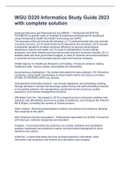
Beat Type Beat characteristics Example
Sinus Normal P wave
Normal (narrow) QRS
Atrial Abnormal P wave
Normal (narrow) QRS
Junctional (AV nodal) P wave absent
(buried in QRS) or;
Abnormal P wave just
before/after QRS
Narrow QRS
Ventricular Wide QRS
Abnormal T wave
No P wave
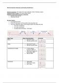
, First degree AV block
Every P wave is followed by a QRS
PR Interval >200ms (>1 big square)
PR interval is constant
Example First degree AV block
Normal conduction: SA node à AV node à Bundle of His à Purkinje system
Disease at the AV node à AV block (heart block)
Disease in the bundle branches or fascicles
Bundle branch blocks
Fascicular blocks
Escape rhythm
If no impulse arrives
If the SA node does not fire another area may take over
The more distal the origin of the escape rhythm the slower the rate
o SA node: 70-80 bpm
o AV node: 40-60 bpm
o Purkinje fibres/ventricles: 15-40 bpm
Beat Type Beat characteristics Example
Sinus Normal P wave
Normal (narrow) QRS
Atrial Abnormal P wave
Normal (narrow) QRS
Junctional (AV nodal) P wave absent
(buried in QRS) or;
Abnormal P wave just
before/after QRS
Narrow QRS
Ventricular Wide QRS
Abnormal T wave
No P wave
, First degree AV block
Every P wave is followed by a QRS
PR Interval >200ms (>1 big square)
PR interval is constant
Example First degree AV block