Table of Contents
ELEMENT 2 – BIOCHEMISTRY ............................................................................................. 1
2.1: Intro to Biochemistry ........................................................................................................... 1
2.2: Protein Folding .................................................................................................................... 2
2.3: Clinical Applications/Separation Techniques........................................................................ 2
2.4, 2.5 & 2.8-2.10: Molecular Genetics ...................................................................................... 3
2.6: Enzymes .............................................................................................................................. 8
2.7: Myoglobin and Haemoglobin ............................................................................................. 10
2.11-2.13: Membranes and Transport ...................................................................................... 10
2.14 – 2.17: Bioenergetics, Fatty Acid Oxidation and the Citrate Cycle ...................................... 15
2.18 & 2.19: Mitochondria and the Electron Transport Chain.................................................... 18
ELEMENT 2 – BIOCHEMISTRY
2.1: Intro to Biochemistry
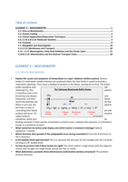
• Explain the causes and symptoms of ketoacidosis in a type I diabetes mellitus patient. Ketone
bodies (3 small water soluble ketones) are produced when the liver thinks it needs to provide a
respiratory substrate. They cause a buildup of protons in the blood, causing pH to drop. The blood
buffer equilibria shift left
releasing CO2. The patient
is therefore seen to be
breathing very deeply and
frequently, and their blood
serum bicarbonate will be low.
When it runs out, the patient
will have no way of
controlling their blood pH and
acidosis will occur. An enzyme
called carbonic
anhydrase, which acts by
binding reactants in their specific orientations so they’ll react, catalyses the reactions. It is therefore
known as a proximity enzyme.
• What isomerism do amino acids display and which isomer is involved in biology? Optical
isomerism. L-isomer.
• Which direction does growth of the polypeptide occur during translation? From the N-terminus to
the C-terminus
• Is the peptide bond rigid and why? Yes it is, because the lone pair on the nitrogen join the C-N bond
forming a C=N+ double bond.
• So how do proteins fold if these bonds are rigid? The chiral carbon’s single bonds with the adjacent
carbon and nitrogen are single bonds and do are free to rotate.
• What determines a proteins three-dimensional conformation (tertiary structure)? The proteins
primary structure.
, 2.2: Protein Folding
• What forces are there stabilising proteins?
1. Hydrogen bonds
2. Ionic bonds (including disulphide bridges) – long range
3. Hydrophobic interactions – hydrophobic amino acids stick together
4. Hydrophilic interactions – hydrophilic amino acids stick together and fold around the
protein, burying the hydrophobic amino acids in the interior of the protein.
• Describe the shape of an alpha helix and the relative positions of the amino acid side chains. A rod
like, right handed helical structure, with ALL of the side chains sticking out of the helix (because all L
optical isomers)
• What is an amino acid residue? Its whats left of the amino acid after peptide bond formation and
subsequent loss of the water molecule. So in alpha amino acid polypeptides its -NH-CHR-COO-
• How is the alpha helix stabilised? Hydrogen bonding between the C=O of one amino acid residue
and the N-H of the amino acid residue four units along (denoted residue i and residue i+4)
• How is the beta pleated sheet stabilised? Through hydrogen bonding between N-H and C=O of
different polypeptide strands, often running in opposite directions, or between the same
polypeptide chain after an abrupt β-turn.
• What group is responsible for disulphide bridges and which amino acid has this group? Thiol,
Cysteine.
• How do proteins span the membrane? Using alpha helices.
• What’s the difference between alpha helices inside the cell membrane and those outside the cell
membrane? Hydrophobic side chains now on the outside of the helix.
2.3: Clinical Applications/Separation Techniques
• How does the gel used in electrophoresis work? Long chains of polyacrylamide with bis-acrylamide
forming cross-links create a mesh-like structure. The varying concentrations of polymer form varying
pore sizes.
• How does gel-electrophoresis separate molecules? When an electric field is applied, molecules
move according to their charge density = overall charge/molecular volume
• How does SDS-PAGE differ? It makes all molecules have the same charge density by changing each
molecules charge proportional to its size. This is done because the SDS evenly coats proteins, making
them all negatively charged, and meaning that the larger molecules have a larger negative charge as
more SDS can attach. Hence charge density is the same.
• What does SDS-PAGE stand for? Sodium dodecyl sulphate polyacrylamide gel electrophoresis
• How are proteins treated before electrophoresis occurs? SDS is added (a reducing agent to remove
disulphide bonds) to the proteins and boiled (to denature them [quaternary structure disappears]
and expose internal hydrophobic sections, onto which the aliphatic carbon dodecyl carbon chain of
the SDS can hydrogen bond to [and since SDS is negatively charged, so too becomes the whole
protein])
• What are isoenzymes? They are enzymes that are the same but are coded for by different genes.
The primary structure is therefore different but the 3D shapes they fold into are very similar. Their
functions are therefore the same but responses are very slightly different (i.e. needs more/less
substrate to function).
• What is ELISA and how does it work? Enzyme linked immunoadsorbent assay – this is the process of
detecting an antigen in a sample. The antibody is associated with a specific enzyme. When the
enzyme reacts with its substrate, a colour change is seen. The process is as follows assuming antigen
is present:
, 1. Antibody with associated enzyme is added to test sample
2. The antibody binds with the antigen
3. Unbounded antibodies are removed, leaving only the antibody/enzyme complexes that are
bound to the antigen
4. The enzymes substrate is added
5. The amount of coloured product formed per unit time is proportional to the amount of
antigen in the substance
It is worth noting that sometimes an extra antibody is involved in the process that binds to the other
antibody. This creates an antibody-antibody-enzyme complex. This is useful when the amount of
antigen you want to test for is very small. This is because in reality it’s an antibody attached to many
other antibodies, each of which has an enzyme attached. So for one antigen present, there’ll be
many enzyme reactions and so more of a colour change.
2.4, 2.5 & 2.8-2.10: Molecular Genetics
• What charge does DNA have and why? Negative because the phosphate in the backbone is
negatively charged due to the –O-.
• In what form do free DNA nucleotides float around in the cell? With a triphosphate onto them
• So what happens when a nucleotide is added onto a growing DNA chain? The outer two
phosphates are cleaved off and are called pyrophosphates (like a little diphosphate). This is split into
two phosphates by a pyrophosphatase enzyme which makes the addition of the nucleotide to the
DNA molecule irreversible.
• Purine .vs. pyrimidine? Double ringed (adenine and guanine) and single ringed (thymine, uracil,
cytosine)
• What can go wrong with cytosine? It can deaminate and turn into uracil, but a special repair
pathway reaminates it.
• Why do bacteria use uracil and not thymine? Because its easier to manufacture
• Why is the deamination of cytosine not a problem in humans? Because in DNA it is repaired through
a special repair pathway and in RNA its not an issue because there are so many mRNA molecules
around.
• Why do humans have thymine instead of uracil? Because if uracil was involved then the body
would think it was a deaminated cytosine and reaminate it.
• What’s a nucleoside? A nucleotide without the phosphate group
• What type of bond links the nitrogenous base to the sugar? Glycosidic bond
• Describe the structure of the DNA helix. A double stranded right handed helix about a common axis.
The strands run in opposite directions and so are said to be antiparallel.
• Describe and explain the relative strengths of the base pairs. A=T is weaker than CΞG because it has
one less hydrogen bond.
• Do you know what the major and minor grooves of the double helix are?
• What is the major grooves function? Allows for more space for the proteins that probe around the
double helix.
• Distinguish between endonucleases and exonucleases. Exonucleases cleave nucleotides one at a
time from the ends of the polynucleotide chain. Endonucleases recognise a restriction site and break
the phosphodiester bond in the backbone there, separating the polynucleotide chain.
• What bond is present in the backbone of a DNA molecule? Phosphodiester bond
• Are most of the DNA polymerases in eukaryotes used for DNA replication or DNA repair? DNA
repair
, • What DNA polymerases in prokaryotes and eukaryotes are used in nuclear DNA replication?
Prokaryotes Eukaryotes
DNA polymerases I & III (mainly III) DNA polymerases α and δ
• What eukaryotic DNA polymerase is used for mitochondrial DNA replication? γ
• What are the four things that DNA polymerase requires in order to function?
1. All 4 nucleotides (deoxyriboadeninetriphosphate [dATP], dGTP, dTTP, dCTP)
2. Mg2+ for the DNA polymerases active site functioning
3. A primer to create a free 3’ end
4. A DNA template strand
• Why is a primer necessary for DNA polymerases functioning during DNA replication? DNA
polymerase can only act on an existing double strand of DNA
• Why can DNA polymerase only act on a double strand of DNA? Because of its proofreading
function, where it looks back on the last nucleotide added to see if it’s correct. If it isn’t then it
cleaves it off using 3’à5’ exonuclease activity and tries again. Without the primer, the DNA
polymerase has nothing to look back on and cannot function.
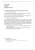
• How do circular chromosomes in prokaryotic
replicate themselves, and what is the theta
structure? Replication starts at the origin of
replication point and proceeds bidirectionally in
opposite directions until two loops appear. The
theta structure is a shape that forms (resembling the
letter theta) approximately halfway into replication.
• What’s DNAa? A protein that initiates DNA
replication in prokaryotes.
• How is the origin of replication different to any other bit of the chromosome/ why does
replication always start there?
1. The bases immediately preceding the origin is rich in A/T linkages, which are easier to break
that G/C linkages because it has one fewer hydrogen bonds
2. There are DNAa protein binding sites
• Approximately how many origin sites are there in humans and why are there so many more than
in prokaryotes? ≈10,000 because otherwise it would take too long
• What is DNAb? It’s the helicase enzyme that uses ATP to unwind DNA
• In the replication fork, how is the fact that DNA polymerase only builds in a 5’à3’ direction
overcome? With the use of discontinuous DNA replication on the lagging strand, leading to the
formation of Okazaki fragments.
• How is the issue of DNA polymerase not being able to synthesise de nova overcome? By using RNA
primase that lays down a small RNA primer.
• Why can RNA primase synthesise de nova but DNA pol cant? Because RNA primase doesn’t have
the same proofreading function that DNA pol has.
• What does DNA polymerase I do?
1. 5’à3’ exonuclease activity - removes RNA nucleotides in the RNA primer ahead of itself
during lagging strand synthesis
2. 5’à3’ synthesis – adds nucleotides during DNA replication on leading strand
3. 3’à5’ proof reading exonuclease activity – chops off nucleotides its just added if it thinks
something has gone wrong (corrects errors during synthesis)
o Note that its only involved with the removal of the RNA primers in lagging strand synthesis
(i.e. it removes RNA primer [function 1], adds a DNA nucleotide [function 2] and then
proofreads what its just added [function 3])
• What does DNA polymerase III do?
a) It’s the main enzyme involved with DNA replication in prokaryotes
b) It never dissociates with the template
ELEMENT 2 – BIOCHEMISTRY ............................................................................................. 1
2.1: Intro to Biochemistry ........................................................................................................... 1
2.2: Protein Folding .................................................................................................................... 2
2.3: Clinical Applications/Separation Techniques........................................................................ 2
2.4, 2.5 & 2.8-2.10: Molecular Genetics ...................................................................................... 3
2.6: Enzymes .............................................................................................................................. 8
2.7: Myoglobin and Haemoglobin ............................................................................................. 10
2.11-2.13: Membranes and Transport ...................................................................................... 10
2.14 – 2.17: Bioenergetics, Fatty Acid Oxidation and the Citrate Cycle ...................................... 15
2.18 & 2.19: Mitochondria and the Electron Transport Chain.................................................... 18
ELEMENT 2 – BIOCHEMISTRY
2.1: Intro to Biochemistry
• Explain the causes and symptoms of ketoacidosis in a type I diabetes mellitus patient. Ketone
bodies (3 small water soluble ketones) are produced when the liver thinks it needs to provide a
respiratory substrate. They cause a buildup of protons in the blood, causing pH to drop. The blood
buffer equilibria shift left
releasing CO2. The patient
is therefore seen to be
breathing very deeply and
frequently, and their blood
serum bicarbonate will be low.
When it runs out, the patient
will have no way of
controlling their blood pH and
acidosis will occur. An enzyme
called carbonic
anhydrase, which acts by
binding reactants in their specific orientations so they’ll react, catalyses the reactions. It is therefore
known as a proximity enzyme.
• What isomerism do amino acids display and which isomer is involved in biology? Optical
isomerism. L-isomer.
• Which direction does growth of the polypeptide occur during translation? From the N-terminus to
the C-terminus
• Is the peptide bond rigid and why? Yes it is, because the lone pair on the nitrogen join the C-N bond
forming a C=N+ double bond.
• So how do proteins fold if these bonds are rigid? The chiral carbon’s single bonds with the adjacent
carbon and nitrogen are single bonds and do are free to rotate.
• What determines a proteins three-dimensional conformation (tertiary structure)? The proteins
primary structure.
, 2.2: Protein Folding
• What forces are there stabilising proteins?
1. Hydrogen bonds
2. Ionic bonds (including disulphide bridges) – long range
3. Hydrophobic interactions – hydrophobic amino acids stick together
4. Hydrophilic interactions – hydrophilic amino acids stick together and fold around the
protein, burying the hydrophobic amino acids in the interior of the protein.
• Describe the shape of an alpha helix and the relative positions of the amino acid side chains. A rod
like, right handed helical structure, with ALL of the side chains sticking out of the helix (because all L
optical isomers)
• What is an amino acid residue? Its whats left of the amino acid after peptide bond formation and
subsequent loss of the water molecule. So in alpha amino acid polypeptides its -NH-CHR-COO-
• How is the alpha helix stabilised? Hydrogen bonding between the C=O of one amino acid residue
and the N-H of the amino acid residue four units along (denoted residue i and residue i+4)
• How is the beta pleated sheet stabilised? Through hydrogen bonding between N-H and C=O of
different polypeptide strands, often running in opposite directions, or between the same
polypeptide chain after an abrupt β-turn.
• What group is responsible for disulphide bridges and which amino acid has this group? Thiol,
Cysteine.
• How do proteins span the membrane? Using alpha helices.
• What’s the difference between alpha helices inside the cell membrane and those outside the cell
membrane? Hydrophobic side chains now on the outside of the helix.
2.3: Clinical Applications/Separation Techniques
• How does the gel used in electrophoresis work? Long chains of polyacrylamide with bis-acrylamide
forming cross-links create a mesh-like structure. The varying concentrations of polymer form varying
pore sizes.
• How does gel-electrophoresis separate molecules? When an electric field is applied, molecules
move according to their charge density = overall charge/molecular volume
• How does SDS-PAGE differ? It makes all molecules have the same charge density by changing each
molecules charge proportional to its size. This is done because the SDS evenly coats proteins, making
them all negatively charged, and meaning that the larger molecules have a larger negative charge as
more SDS can attach. Hence charge density is the same.
• What does SDS-PAGE stand for? Sodium dodecyl sulphate polyacrylamide gel electrophoresis
• How are proteins treated before electrophoresis occurs? SDS is added (a reducing agent to remove
disulphide bonds) to the proteins and boiled (to denature them [quaternary structure disappears]
and expose internal hydrophobic sections, onto which the aliphatic carbon dodecyl carbon chain of
the SDS can hydrogen bond to [and since SDS is negatively charged, so too becomes the whole
protein])
• What are isoenzymes? They are enzymes that are the same but are coded for by different genes.
The primary structure is therefore different but the 3D shapes they fold into are very similar. Their
functions are therefore the same but responses are very slightly different (i.e. needs more/less
substrate to function).
• What is ELISA and how does it work? Enzyme linked immunoadsorbent assay – this is the process of
detecting an antigen in a sample. The antibody is associated with a specific enzyme. When the
enzyme reacts with its substrate, a colour change is seen. The process is as follows assuming antigen
is present:
, 1. Antibody with associated enzyme is added to test sample
2. The antibody binds with the antigen
3. Unbounded antibodies are removed, leaving only the antibody/enzyme complexes that are
bound to the antigen
4. The enzymes substrate is added
5. The amount of coloured product formed per unit time is proportional to the amount of
antigen in the substance
It is worth noting that sometimes an extra antibody is involved in the process that binds to the other
antibody. This creates an antibody-antibody-enzyme complex. This is useful when the amount of
antigen you want to test for is very small. This is because in reality it’s an antibody attached to many
other antibodies, each of which has an enzyme attached. So for one antigen present, there’ll be
many enzyme reactions and so more of a colour change.
2.4, 2.5 & 2.8-2.10: Molecular Genetics
• What charge does DNA have and why? Negative because the phosphate in the backbone is
negatively charged due to the –O-.
• In what form do free DNA nucleotides float around in the cell? With a triphosphate onto them
• So what happens when a nucleotide is added onto a growing DNA chain? The outer two
phosphates are cleaved off and are called pyrophosphates (like a little diphosphate). This is split into
two phosphates by a pyrophosphatase enzyme which makes the addition of the nucleotide to the
DNA molecule irreversible.
• Purine .vs. pyrimidine? Double ringed (adenine and guanine) and single ringed (thymine, uracil,
cytosine)
• What can go wrong with cytosine? It can deaminate and turn into uracil, but a special repair
pathway reaminates it.
• Why do bacteria use uracil and not thymine? Because its easier to manufacture
• Why is the deamination of cytosine not a problem in humans? Because in DNA it is repaired through
a special repair pathway and in RNA its not an issue because there are so many mRNA molecules
around.
• Why do humans have thymine instead of uracil? Because if uracil was involved then the body
would think it was a deaminated cytosine and reaminate it.
• What’s a nucleoside? A nucleotide without the phosphate group
• What type of bond links the nitrogenous base to the sugar? Glycosidic bond
• Describe the structure of the DNA helix. A double stranded right handed helix about a common axis.
The strands run in opposite directions and so are said to be antiparallel.
• Describe and explain the relative strengths of the base pairs. A=T is weaker than CΞG because it has
one less hydrogen bond.
• Do you know what the major and minor grooves of the double helix are?
• What is the major grooves function? Allows for more space for the proteins that probe around the
double helix.
• Distinguish between endonucleases and exonucleases. Exonucleases cleave nucleotides one at a
time from the ends of the polynucleotide chain. Endonucleases recognise a restriction site and break
the phosphodiester bond in the backbone there, separating the polynucleotide chain.
• What bond is present in the backbone of a DNA molecule? Phosphodiester bond
• Are most of the DNA polymerases in eukaryotes used for DNA replication or DNA repair? DNA
repair
, • What DNA polymerases in prokaryotes and eukaryotes are used in nuclear DNA replication?
Prokaryotes Eukaryotes
DNA polymerases I & III (mainly III) DNA polymerases α and δ
• What eukaryotic DNA polymerase is used for mitochondrial DNA replication? γ
• What are the four things that DNA polymerase requires in order to function?
1. All 4 nucleotides (deoxyriboadeninetriphosphate [dATP], dGTP, dTTP, dCTP)
2. Mg2+ for the DNA polymerases active site functioning
3. A primer to create a free 3’ end
4. A DNA template strand
• Why is a primer necessary for DNA polymerases functioning during DNA replication? DNA
polymerase can only act on an existing double strand of DNA
• Why can DNA polymerase only act on a double strand of DNA? Because of its proofreading
function, where it looks back on the last nucleotide added to see if it’s correct. If it isn’t then it
cleaves it off using 3’à5’ exonuclease activity and tries again. Without the primer, the DNA
polymerase has nothing to look back on and cannot function.
• How do circular chromosomes in prokaryotic
replicate themselves, and what is the theta
structure? Replication starts at the origin of
replication point and proceeds bidirectionally in
opposite directions until two loops appear. The
theta structure is a shape that forms (resembling the
letter theta) approximately halfway into replication.
• What’s DNAa? A protein that initiates DNA
replication in prokaryotes.
• How is the origin of replication different to any other bit of the chromosome/ why does
replication always start there?
1. The bases immediately preceding the origin is rich in A/T linkages, which are easier to break
that G/C linkages because it has one fewer hydrogen bonds
2. There are DNAa protein binding sites
• Approximately how many origin sites are there in humans and why are there so many more than
in prokaryotes? ≈10,000 because otherwise it would take too long
• What is DNAb? It’s the helicase enzyme that uses ATP to unwind DNA
• In the replication fork, how is the fact that DNA polymerase only builds in a 5’à3’ direction
overcome? With the use of discontinuous DNA replication on the lagging strand, leading to the
formation of Okazaki fragments.
• How is the issue of DNA polymerase not being able to synthesise de nova overcome? By using RNA
primase that lays down a small RNA primer.
• Why can RNA primase synthesise de nova but DNA pol cant? Because RNA primase doesn’t have
the same proofreading function that DNA pol has.
• What does DNA polymerase I do?
1. 5’à3’ exonuclease activity - removes RNA nucleotides in the RNA primer ahead of itself
during lagging strand synthesis
2. 5’à3’ synthesis – adds nucleotides during DNA replication on leading strand
3. 3’à5’ proof reading exonuclease activity – chops off nucleotides its just added if it thinks
something has gone wrong (corrects errors during synthesis)
o Note that its only involved with the removal of the RNA primers in lagging strand synthesis
(i.e. it removes RNA primer [function 1], adds a DNA nucleotide [function 2] and then
proofreads what its just added [function 3])
• What does DNA polymerase III do?
a) It’s the main enzyme involved with DNA replication in prokaryotes
b) It never dissociates with the template