GEN2103 – Lectures
Anatomy of the brain and histology
- Neuroscience is important because it is an emerging field, because all the functions
of your body is controlled by your brain. There are neurological diseases like MS
(multiple sclerosis) and depression. Aging is a very important factor in diseases, also
genes and environment will influence the brain.
Microscopic anatomy
- One trillion (10^12) nerve cells linked together to form the rapid control center of
the body. The nerve cells must be linked together to get this rapid action.
- The higher you go in the vertebrae, you’ll see a bigger growth in the forebrain (this is
related to memory formation, decision making and cognition) and you’ll see a
difference between sulci and gyri.
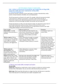
- General organization, the nervous system this is compromised of the central nervous
system and the peripheral nervous system. The peripheral nervous system can be
divided in the autonomic and the somatic part. For the autonomic part we have the
afferent and efferent nerves, the efferent nerves can be divided in the sympatic and
parasympathic nerves. The somatic nervous system can also be divided in afferent
(motoric) and efferent nerves (sensory).
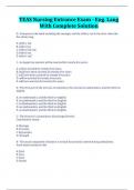
- The CNS can be divided in the brain, which consists of the cerebrum (the forebrain),
the brainstem and the cerebellum (small brain on top of the brain stem). In the
brainstem you’ll find the key areas which contain cells which are vital for the brain
(dopamine in substantia nigra) you’ll have the serotonergic cells (5-HT cells)
(depression) and the noradrenergic cells (epilepsy, Alzheimer’s).
- We have two parts of the brain which are separated from each other and connected
by the corpus callosum. The forebrain can be divided by the telencephalon and
diencephalon. The diencephalon can also be determined as the thalamus, the major
function is in the endocrine system with the hypothalamus. The midbrain
(mesencephalon) and the hindbrain which is divided by the metencephalon and the
myelencephalon. These two form the brainstem. The midbrain is for a major part the
hippocampus. The hindbrain will transfer information from and to the spinal cord.
- Brainstem cancer is the most common form of cancer in the brain. It is always
related to the hindbrain and sometimes in the midbrain.
- Terminology, this is important because you’ll have to communicate with surgeons
where you want DBS to take place. The upper part of the brain is the superior or
dorsal part. De bottom is the inferior or ventral part. Then you have anterior (rostral)
for the front and posterior (caudal) for the back.
,- If you make sections to the brain, we can make different sections: Coronal section
(frontal section), Horizontal section, mid-sagittal section (middle of the brain).
- Mammillary bodies make histamine and are involved in making memories.
- Subcortical structures if you do a mid-sagittal clift you can recognize the corpus
callosum. This contains a lot of white matter (glia cells). Then you can see the
hippocampus and at the end of this the amygdala. The amygdala regulates emotion
and because the amygdala and hippocampus are so closely related emotion and
memory are linked together.
- Medial there are other glands to find, the pineal gland (epiphysis – produces
melatonin which is important for sleep), white matter on top of the thalamus which
is the fornix, tegmentum, and tectum.
- The outside of the brain, we have three areas: the dura mater between the brain and
skull, the arachnoid membrane, and the pia mater this is on top of the brain. The
dura is composed of two layers, this is to keep the brain stable it is tough and
flexible. It surrounds the brain and the spinal cord. The sub-arachnoid space is the
space in which very small blood vessels are located. Toxic products from the brain
grey matter moves down the spinal cord via this and goes down to the kidneys.
- We have gyri which are the top levels, then smaller holes which we call sulci and
then the big ones we call fissures. One fissure is the division between temporal lobe
and the parietal lobe. We also have a fissure which separates the motor area with
the sensory area.
- In the Alzheimer brain we’ll see shrinkage of the brain. The cells are getting closer
because of the loss of water. In the Alzheimer brain there are the same number of
neurons, but they are getting closer to each other. This will lead to enlarged
ventricles, shrinkage of the language area, the increase of big sulci and small gyri,
also the hippocampus shrunk which leads to loss of memory.
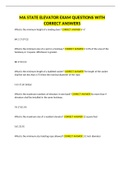
- We have several lobes, the occipital lobe, the parietal lobe, and the frontal lobe.
Between the parietal lobe and frontal lobe, we see the central sulcus. Then we have
the lateral fissure between the temporal lobe and other lobes. The function of the
lobes are as follows: Parietal lobe motor behavior, occipital lobe with vision and
vision processes. The frontal lobe with cognition. And the temporal lobe with
learning.
- Wernicke area is involved with speaking in the parietal lobe. The most important
part is the prefrontal cortex which is on top of the premotor area. Broca’s area is for
motor speech area, it is in the ventral part of the premotor cortex (control of mouth,
muscles etc). Wernicke’s area is related to speech and in the parietal lobe.
- The frontal lobe is divided by two fissures it is separated from the temporal lobe by
the lateral fissure. The central sulcus separates the frontal lobe from the parietal
lobe. The functions of this lobe are the higher cognitive functions. It is related also
with memory, language, motivation, problem solving, decision making and
personality. When you have Alzheimer’s, you’ll lose these functions.
- The parietal lobe is separated by the central sulcus, and by the lateral fissure. It is
also separated from the occipital lobe by the parieto-occipital sulcus. It is mostly for
sensory processing (smell, taste, touch), it has to do with sensory and spatial
recognition and lastly with eye-hand coordination.
- The temporal lobe it is separated from the upper two by the lateral fissure, and from
the occipital lobe by the occipital sulcus. It is related to two important brain
, structures, which makes it important for memory and emotions. Sexual behavior,
aggression etc are related to this lobe.
- The occipital lobe is strictly related to view, and eyesight. It is separated by the
parietal-occipital sulcus and the occipital sulcus.
- The 5th lobe is the insula cortex, which is under the temporal lobe. When you open
the brain, you can see inside the brain and see the cortex, this is the insula cortex.
The exact role is not yet understood.
- When you look from dorsal to the brain, you can see the white matter (corpus
callosum) as the bridge between the hemispheres. Then you have the commisura
anterior (in the front) and the commisura posterior (in the back).
- Ventricle systems are the places in the brain where fluid is available, they are
important for imaging because the ventricles are the first thing you’ll look at. When
they are enlarged, you’ll know where the problem is.
- The biggest are the lateral ventricles, these are situated in the telencephalon. The
third ventricle is in the diencephalon. Under in the cerebellum we have the fourth
ventricle. There is a cerebral aqueduct in the metencephalon which is the connection
between 3rd and 4th ventricle. The ventricular system is important for protection and
transport of fluid and toxins.
- You’ll go to collect the CSF in the lower part of the lumbar system. This way you can
evade the nerves. This is at lumbar L2, L3, L4, here you can collect a couple of drops
of CSF. The subarachnoid space will collect the waste of the brain, where it will be
transferred down to the spinal cord. This way the CSF can be examined.
- The fluid always flows down to the spinal cord, where it eventually mixes with
arterial blood where the toxins can be exchanged, and the kidneys can work their
job. Toxins will be transferred to the subarachnoid space and then transferred down
to the spinal fluid.
- The caudate-putamen complex, this area of the brain is related to motor functions.
Whatever you want to do with your muscles. Your brain must give your body a
direction. That is basically an effect starting in the caudate nucleus, which goes on to
the spinal cord eventually.
- Normally when you want to flee, your brain will look for a response you’ve done in
the past. If that is a good strategy the information will be relayed to the caudate
nucleus, which will send its information to the putamen in the correct part to
process it.
- Limbic system is one of the most important systems. The limbic system has to do
with learning. The limbic system starts at the mammillary bodies these send fibers to
the hypothalamus; these are located as bundles in the fornix. Under the corpus
callosum and above the thalamus. Towards the hippocampus. It is involved in
emotions, motivation, and emotional association with memory. Also, depression can
start here.
Limbic system also regulates the hypothalamus, is involved with regulation of
emotion and learning and memories.
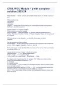
- The circuit of Papez means that information from the cortex which can go into the
cingulate gyrus which processes information to the hippocampus, towards the
mammillary bodies which can activate the thalamus particularly the anterior nucleus
of the thalamus towards the cingulate gyrus. But most of the times the hippocampus
will activate the hypothalamus which sends the signal back to the cortex.
Anatomy of the brain and histology
- Neuroscience is important because it is an emerging field, because all the functions
of your body is controlled by your brain. There are neurological diseases like MS
(multiple sclerosis) and depression. Aging is a very important factor in diseases, also
genes and environment will influence the brain.
Microscopic anatomy
- One trillion (10^12) nerve cells linked together to form the rapid control center of
the body. The nerve cells must be linked together to get this rapid action.
- The higher you go in the vertebrae, you’ll see a bigger growth in the forebrain (this is
related to memory formation, decision making and cognition) and you’ll see a
difference between sulci and gyri.
- General organization, the nervous system this is compromised of the central nervous
system and the peripheral nervous system. The peripheral nervous system can be
divided in the autonomic and the somatic part. For the autonomic part we have the
afferent and efferent nerves, the efferent nerves can be divided in the sympatic and
parasympathic nerves. The somatic nervous system can also be divided in afferent
(motoric) and efferent nerves (sensory).
- The CNS can be divided in the brain, which consists of the cerebrum (the forebrain),
the brainstem and the cerebellum (small brain on top of the brain stem). In the
brainstem you’ll find the key areas which contain cells which are vital for the brain
(dopamine in substantia nigra) you’ll have the serotonergic cells (5-HT cells)
(depression) and the noradrenergic cells (epilepsy, Alzheimer’s).
- We have two parts of the brain which are separated from each other and connected
by the corpus callosum. The forebrain can be divided by the telencephalon and
diencephalon. The diencephalon can also be determined as the thalamus, the major
function is in the endocrine system with the hypothalamus. The midbrain
(mesencephalon) and the hindbrain which is divided by the metencephalon and the
myelencephalon. These two form the brainstem. The midbrain is for a major part the
hippocampus. The hindbrain will transfer information from and to the spinal cord.
- Brainstem cancer is the most common form of cancer in the brain. It is always
related to the hindbrain and sometimes in the midbrain.
- Terminology, this is important because you’ll have to communicate with surgeons
where you want DBS to take place. The upper part of the brain is the superior or
dorsal part. De bottom is the inferior or ventral part. Then you have anterior (rostral)
for the front and posterior (caudal) for the back.
,- If you make sections to the brain, we can make different sections: Coronal section
(frontal section), Horizontal section, mid-sagittal section (middle of the brain).
- Mammillary bodies make histamine and are involved in making memories.
- Subcortical structures if you do a mid-sagittal clift you can recognize the corpus
callosum. This contains a lot of white matter (glia cells). Then you can see the
hippocampus and at the end of this the amygdala. The amygdala regulates emotion
and because the amygdala and hippocampus are so closely related emotion and
memory are linked together.
- Medial there are other glands to find, the pineal gland (epiphysis – produces
melatonin which is important for sleep), white matter on top of the thalamus which
is the fornix, tegmentum, and tectum.
- The outside of the brain, we have three areas: the dura mater between the brain and
skull, the arachnoid membrane, and the pia mater this is on top of the brain. The
dura is composed of two layers, this is to keep the brain stable it is tough and
flexible. It surrounds the brain and the spinal cord. The sub-arachnoid space is the
space in which very small blood vessels are located. Toxic products from the brain
grey matter moves down the spinal cord via this and goes down to the kidneys.
- We have gyri which are the top levels, then smaller holes which we call sulci and
then the big ones we call fissures. One fissure is the division between temporal lobe
and the parietal lobe. We also have a fissure which separates the motor area with
the sensory area.
- In the Alzheimer brain we’ll see shrinkage of the brain. The cells are getting closer
because of the loss of water. In the Alzheimer brain there are the same number of
neurons, but they are getting closer to each other. This will lead to enlarged
ventricles, shrinkage of the language area, the increase of big sulci and small gyri,
also the hippocampus shrunk which leads to loss of memory.
- We have several lobes, the occipital lobe, the parietal lobe, and the frontal lobe.
Between the parietal lobe and frontal lobe, we see the central sulcus. Then we have
the lateral fissure between the temporal lobe and other lobes. The function of the
lobes are as follows: Parietal lobe motor behavior, occipital lobe with vision and
vision processes. The frontal lobe with cognition. And the temporal lobe with
learning.
- Wernicke area is involved with speaking in the parietal lobe. The most important
part is the prefrontal cortex which is on top of the premotor area. Broca’s area is for
motor speech area, it is in the ventral part of the premotor cortex (control of mouth,
muscles etc). Wernicke’s area is related to speech and in the parietal lobe.
- The frontal lobe is divided by two fissures it is separated from the temporal lobe by
the lateral fissure. The central sulcus separates the frontal lobe from the parietal
lobe. The functions of this lobe are the higher cognitive functions. It is related also
with memory, language, motivation, problem solving, decision making and
personality. When you have Alzheimer’s, you’ll lose these functions.
- The parietal lobe is separated by the central sulcus, and by the lateral fissure. It is
also separated from the occipital lobe by the parieto-occipital sulcus. It is mostly for
sensory processing (smell, taste, touch), it has to do with sensory and spatial
recognition and lastly with eye-hand coordination.
- The temporal lobe it is separated from the upper two by the lateral fissure, and from
the occipital lobe by the occipital sulcus. It is related to two important brain
, structures, which makes it important for memory and emotions. Sexual behavior,
aggression etc are related to this lobe.
- The occipital lobe is strictly related to view, and eyesight. It is separated by the
parietal-occipital sulcus and the occipital sulcus.
- The 5th lobe is the insula cortex, which is under the temporal lobe. When you open
the brain, you can see inside the brain and see the cortex, this is the insula cortex.
The exact role is not yet understood.
- When you look from dorsal to the brain, you can see the white matter (corpus
callosum) as the bridge between the hemispheres. Then you have the commisura
anterior (in the front) and the commisura posterior (in the back).
- Ventricle systems are the places in the brain where fluid is available, they are
important for imaging because the ventricles are the first thing you’ll look at. When
they are enlarged, you’ll know where the problem is.
- The biggest are the lateral ventricles, these are situated in the telencephalon. The
third ventricle is in the diencephalon. Under in the cerebellum we have the fourth
ventricle. There is a cerebral aqueduct in the metencephalon which is the connection
between 3rd and 4th ventricle. The ventricular system is important for protection and
transport of fluid and toxins.
- You’ll go to collect the CSF in the lower part of the lumbar system. This way you can
evade the nerves. This is at lumbar L2, L3, L4, here you can collect a couple of drops
of CSF. The subarachnoid space will collect the waste of the brain, where it will be
transferred down to the spinal cord. This way the CSF can be examined.
- The fluid always flows down to the spinal cord, where it eventually mixes with
arterial blood where the toxins can be exchanged, and the kidneys can work their
job. Toxins will be transferred to the subarachnoid space and then transferred down
to the spinal fluid.
- The caudate-putamen complex, this area of the brain is related to motor functions.
Whatever you want to do with your muscles. Your brain must give your body a
direction. That is basically an effect starting in the caudate nucleus, which goes on to
the spinal cord eventually.
- Normally when you want to flee, your brain will look for a response you’ve done in
the past. If that is a good strategy the information will be relayed to the caudate
nucleus, which will send its information to the putamen in the correct part to
process it.
- Limbic system is one of the most important systems. The limbic system has to do
with learning. The limbic system starts at the mammillary bodies these send fibers to
the hypothalamus; these are located as bundles in the fornix. Under the corpus
callosum and above the thalamus. Towards the hippocampus. It is involved in
emotions, motivation, and emotional association with memory. Also, depression can
start here.
Limbic system also regulates the hypothalamus, is involved with regulation of
emotion and learning and memories.
- The circuit of Papez means that information from the cortex which can go into the
cingulate gyrus which processes information to the hippocampus, towards the
mammillary bodies which can activate the thalamus particularly the anterior nucleus
of the thalamus towards the cingulate gyrus. But most of the times the hippocampus
will activate the hypothalamus which sends the signal back to the cortex.