University of Cambridge
MVST Part IA Molecules in Medical Sciences
Biological macromolecules, protein structure and enzyme catalysis
Dr Helen Mott
Lecture 1:
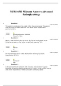
• Glucose = aldose at C1, 4 chiral centres (C2, C3, C4, C5), Beta chair=2 conformations
• Monosaccharides more than 5C usually cyclic > new chiral centre > α- and β-ring
enantiomers
o fructose = ketose at C2
• Free end of chain = reducing end > ring can be opened to produce free reducing aldehyde
group
• Glycogen more ends to cut glucose – accessible – short term store – water between units
• Cellulose – beta 1,4 alternate aspect > very straight chained > held by H-bonds to form
microfibrils
• Chitin = N-acetyl glucosamine.
• Oligosaccharides: N-Asp linked or O-Ser/Thre linked > added ER > processed Golgi > on cell
surface
• DNA sugar: Beta-2-deoxyribose = aldopentose
• Nucleoside = ribose + base > nucleotide = ribose + base + phosphate > phosphoanhydride
bonds = energy
• DNA helix turns every 3.4nm/10 nucleotides
• tRNA: single-stranded but some areas complementary > hairpin loops > 4 arms
• AA: alpha carbon > L-form
• Hydrophobic aliphatic: cluster away from water, pack tightly: ALVPIG
o Proline: rigid ring > bends and kinks. No NH2 = no H-bond
• Charged: ionic interactions: acid/base catalysis: +ve= R(Arginine), K(Lysine), H > -ve=
D(Aspartic Acid), E(Glutamic acid),
,• Polar: H=bonds on surface: N(Asparagine), Q(Glutamine), S, T, Y
• Aromatic: hydrophobic: F(Phe), W(Trytophan)
• Sulphur containing: C, M(hydrophobic)
➢ Peptide bond planar due to electron delocalisation > free rotation around alpha carbon >
folding occurs by rotation of the φ and ψ angles.
Lecture 2: Protein 3D structure
Primary structure (1°) = linear sequence of AA > determines overall structure
• Experiments: Sample RNAase was unfolded/denatured in test tube by adding urea
(disrupts the non-covalent forces) and mercaptoethanol (reduces disulphide bonds) >
denaturing agents removed > spontaneously refolded > active = native structure had re-
formed.
➢ Secondary structure (2°) = folding of regions into localised, regular arrangements of
backbone > due to H-bonds between N-H and C=O > alpha helices or beta pleated sheets.
Lecture 3: Protein structure vs function
➢ Co-factors: reactivity not in AA side chains, from vitamin and minerals, carrying e- or O2, co-
enzymes
➢ Prosthetic group: tightly bound, required for structure e.g. haem, FAD (riboflavin)
• Oxygen binds to central Fe2+
• Carry e- in Fe2+/Fe3+
➢ Co-substrate: used once, released and regenerated e.g. NAD+ (niacin), Co-A
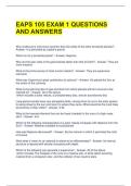
➢ Hb, Mb: hydrophobic pocket for correct geometrical haem binding
• Mb: 153 AA, 8 a-helices > hyperbolic saturation > diffusion within muscle tissues
➢ Hb: tetramer with 2 alpha and 2 beta subunits. Sigmoidal cooperativity
• Flexible: held together by weak interactions that break and reform
• Deoxy: porphyrin ring dome, Fe above ring
• Oxy: Fe in plane of ring, pulls His down and F helix connected to FG loop at interface
> reform H-bonds as subunits slide past each other
• Both: Small 3° changes within subunits lead to strain at interface > concerted
transition
, ➢ Cooperativity: different subunits affect each other’s function
• Symmetry: all subunits R or T, individual subunits can’t change independently >
change conform after first binding
• Sequential: strain at interface when one subunit changes conform > symmetry not
preserved
➢ Membrane proteins: hydrophobic AA outside made of helices and B-barrels > all C=O and N-
H form H-bonds > neutralise polarity inside the membrane > energetically favourable to
forms bonds within chain
• Hydrophilic inside: allow transfer of polar substances
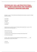
➢ Potassium channel: tetramer identical helical subunits (each subunit contains 3 alpha
helices)
• -ve AA > repel Cl- at top and bottom
• Selectivity filter: strip aqueous shell and provide C=O oxygen mimic hydration
• Na+ too small to contact C=O, stays hydrated so too big
• Closed: hydrophobic side chain blocks pore > Helix rotation at the cytoplasmic face
opens channel
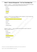
➢ Antibodies: 12 domains, tetramer: 2 identical light and heavy chains, disulphide bonds
(intramolecular, tertiary)
• Each chain has variable Ig domain at N-terminus. VH and VL form antigen binding site
• Extra loops=complementary determining regions (CDRs), rest B-sheet
• CDRs determine specificity, 3 on each chain > recombination
• Light chain has one constant domain = CL binds to CH1
• CH2, CH3(Fc) = dimerization + interact with receptors, different effector functions
MVST Part IA Molecules in Medical Sciences
Biological macromolecules, protein structure and enzyme catalysis
Dr Helen Mott
Lecture 1:
• Glucose = aldose at C1, 4 chiral centres (C2, C3, C4, C5), Beta chair=2 conformations
• Monosaccharides more than 5C usually cyclic > new chiral centre > α- and β-ring
enantiomers
o fructose = ketose at C2
• Free end of chain = reducing end > ring can be opened to produce free reducing aldehyde
group
• Glycogen more ends to cut glucose – accessible – short term store – water between units
• Cellulose – beta 1,4 alternate aspect > very straight chained > held by H-bonds to form
microfibrils
• Chitin = N-acetyl glucosamine.
• Oligosaccharides: N-Asp linked or O-Ser/Thre linked > added ER > processed Golgi > on cell
surface
• DNA sugar: Beta-2-deoxyribose = aldopentose
• Nucleoside = ribose + base > nucleotide = ribose + base + phosphate > phosphoanhydride
bonds = energy
• DNA helix turns every 3.4nm/10 nucleotides
• tRNA: single-stranded but some areas complementary > hairpin loops > 4 arms
• AA: alpha carbon > L-form
• Hydrophobic aliphatic: cluster away from water, pack tightly: ALVPIG
o Proline: rigid ring > bends and kinks. No NH2 = no H-bond
• Charged: ionic interactions: acid/base catalysis: +ve= R(Arginine), K(Lysine), H > -ve=
D(Aspartic Acid), E(Glutamic acid),
,• Polar: H=bonds on surface: N(Asparagine), Q(Glutamine), S, T, Y
• Aromatic: hydrophobic: F(Phe), W(Trytophan)
• Sulphur containing: C, M(hydrophobic)
➢ Peptide bond planar due to electron delocalisation > free rotation around alpha carbon >
folding occurs by rotation of the φ and ψ angles.
Lecture 2: Protein 3D structure
Primary structure (1°) = linear sequence of AA > determines overall structure
• Experiments: Sample RNAase was unfolded/denatured in test tube by adding urea
(disrupts the non-covalent forces) and mercaptoethanol (reduces disulphide bonds) >
denaturing agents removed > spontaneously refolded > active = native structure had re-
formed.
➢ Secondary structure (2°) = folding of regions into localised, regular arrangements of
backbone > due to H-bonds between N-H and C=O > alpha helices or beta pleated sheets.
Lecture 3: Protein structure vs function
➢ Co-factors: reactivity not in AA side chains, from vitamin and minerals, carrying e- or O2, co-
enzymes
➢ Prosthetic group: tightly bound, required for structure e.g. haem, FAD (riboflavin)
• Oxygen binds to central Fe2+
• Carry e- in Fe2+/Fe3+
➢ Co-substrate: used once, released and regenerated e.g. NAD+ (niacin), Co-A
➢ Hb, Mb: hydrophobic pocket for correct geometrical haem binding
• Mb: 153 AA, 8 a-helices > hyperbolic saturation > diffusion within muscle tissues
➢ Hb: tetramer with 2 alpha and 2 beta subunits. Sigmoidal cooperativity
• Flexible: held together by weak interactions that break and reform
• Deoxy: porphyrin ring dome, Fe above ring
• Oxy: Fe in plane of ring, pulls His down and F helix connected to FG loop at interface
> reform H-bonds as subunits slide past each other
• Both: Small 3° changes within subunits lead to strain at interface > concerted
transition
, ➢ Cooperativity: different subunits affect each other’s function
• Symmetry: all subunits R or T, individual subunits can’t change independently >
change conform after first binding
• Sequential: strain at interface when one subunit changes conform > symmetry not
preserved
➢ Membrane proteins: hydrophobic AA outside made of helices and B-barrels > all C=O and N-
H form H-bonds > neutralise polarity inside the membrane > energetically favourable to
forms bonds within chain
• Hydrophilic inside: allow transfer of polar substances
➢ Potassium channel: tetramer identical helical subunits (each subunit contains 3 alpha
helices)
• -ve AA > repel Cl- at top and bottom
• Selectivity filter: strip aqueous shell and provide C=O oxygen mimic hydration
• Na+ too small to contact C=O, stays hydrated so too big
• Closed: hydrophobic side chain blocks pore > Helix rotation at the cytoplasmic face
opens channel
➢ Antibodies: 12 domains, tetramer: 2 identical light and heavy chains, disulphide bonds
(intramolecular, tertiary)
• Each chain has variable Ig domain at N-terminus. VH and VL form antigen binding site
• Extra loops=complementary determining regions (CDRs), rest B-sheet
• CDRs determine specificity, 3 on each chain > recombination
• Light chain has one constant domain = CL binds to CH1
• CH2, CH3(Fc) = dimerization + interact with receptors, different effector functions