CHROMOSOMES
Cellular organisation of genetic material
● All DNA in a cell is the cells genome
○ The genome can consist of a single DNA molecule of many DNA
molecules
■ Prokaryotic cells generally only have a single, circular molecule
making up the genome
■ Eukaryotic organisms have a number of DNA molecules
● DNA are associated with proteins (histones in eukaryotes) that package the DNA
● Chromosome: a single piece of DNA associated with packing proteins
○ DNA interacts with certain dyes
○ When positively charged dyes are added to DNA (which is negatively
charged), strong interactions occur
● 2m of DNA is contained within one cell
○ This is made possible by the very tight packing of DNA in chromosomes
■ As well as the fact that DNA is very thin
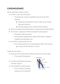
Condensed chromosomes
● During cell division chromosomes condense to become more clearly visible
○ (This is after DNA replication)
● The sister chromatids have exactly the
same DNA sequence
● The end regions are called telomeres
○ They are made up of repetitive
sequences of non coding DNA
, ○ Recall DNA replication: the primer is added before DNA pol III can add
nucleotides however after these primers are removed by DNA pol I, there
is just empty space at the end of the DNA strand
■ The longer strand therefore shortens so it can be the same length
as the replicated strand
○ Telomerase act as a buffer to delay gene shortening - telomerase is an
enzyme that extends the telomeres
■ Telomerase uses repeated sequences to do this
○ Human germ cells contain telomerase to ensure that DNA is not lost from
one generation to another
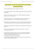
The types of chromosomes
Based on the position of the centromere
● Note that telocentric chromosomes are not found in human cells
Chromosomes in human cells
● Somatic cells (not sex cells) have 23 pairs of chromosomes
, ○ 22 pairs of chromosomes and 1 pair of sex chromosomes
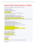
● Karyotype: an ordered display of the pairs of chromosomes from a cell
○ Chromosomes are ordered according to size and staining pattern, position
of chromosome
■ Note that the staining pattern comes about from the way that the
chromosomes interact with certain dyes - different genes interact
differently with different dyes
● Why are chromosomes paired? Every person has a set of chromosomes from their
mother and another set from their father
● Human chromosomes are grouped into 7 classes A-G according to length and
position of centromere
You don’t need to know this table
, ● Two chromosomes in each pair are homologous chromosomes. One chromosome
is a homolog of another
○ They carry genes that control the same characters (e.g both have genes
that code for eye colour, or both have genes that code for the enzyme
helicase)
○ The genes occur at the same position on each homologous pair.
● The sex chromosomes are X and Y
○ Note that the sex chromosomes in males (X and Y) are not homologous
Preparation of a karyotype
● Somatic cells are harvested and grown in a culture
● They are stimulated to divide - this is important at because the condensed form of
chromosomes only occurs in dividing cells
● The division process is stopped during metaphase - they are in their most highly
condensed form at this phase
● The chromosomes are stained and a photograph is taken
○ The most common staining technique is the Giemsa stain which produces
the banding patterns on chromosomes
■ The bands are called G bands
■ Dark stained areas are rich in A and T, called heterochromatic
regions. These are the inactive chromosomal regions (tightly
packed)
■ Light stained regions are euchromatic - they are associated with
active genes (loosely packed)
,LARGE SCALE CHROMOSOMAL CHANGES
● Some genetic disorders occur as a result of alterations of the chromosome
number or structure
● The karyotype can be used to detect abnormal changes
○ Having missing or extra chromosomes is called aneuploidy
● Almost all changes in the chromosome number or structure are so disastrous to
the developing organism that it will immediately abort
○ This is because the lack of / addition of certain genes can upset the genetic
balance
● Some organisms are able to survive with this genetic imbalance but they will
have a syndrome that is characteristic of that aneuploidy
Down syndrome
● Trisomy 21
● It is an aneuploid condition that results in three copies of chromosome 21 (instead
of 2)
● The extra copy affects the organism but not so much that it dies
● it is different from the trisomy of other autosomes in that children can survive into
adulthood
○ Trisomy 18 is Edwards Syndrome
○ Trisomy 13 is Patau syndrome
■ Humans with these syndromes do not survive long after birth
Aneuploidy of sex chromosomes
● These do not affect genetic balance as much as changes in autosomes
● XXX chromosomes - have no unusual physical features
● Monosomy X - produces sterile females
, ● XXY individuals have Klinefelter syndrome - usually does not become noticeable
until adulthood
○ They have a low sperm count
How genetic imbalance occurs
● Duplicating a single gene usually does not have severe consequences
● But duplicating (or removing) ALL the genes on a chromosome does - there are
too many genes in the organism
○ That is why offspring with aneuploidy are not viable
X inactivation in Female mammals
● All humans operate with at least one X chromosome
● One of the X chromosomes in human females is randomly inactivated during
embryonic development
Cellular organisation of genetic material
● All DNA in a cell is the cells genome
○ The genome can consist of a single DNA molecule of many DNA
molecules
■ Prokaryotic cells generally only have a single, circular molecule
making up the genome
■ Eukaryotic organisms have a number of DNA molecules
● DNA are associated with proteins (histones in eukaryotes) that package the DNA
● Chromosome: a single piece of DNA associated with packing proteins
○ DNA interacts with certain dyes
○ When positively charged dyes are added to DNA (which is negatively
charged), strong interactions occur
● 2m of DNA is contained within one cell
○ This is made possible by the very tight packing of DNA in chromosomes
■ As well as the fact that DNA is very thin
Condensed chromosomes
● During cell division chromosomes condense to become more clearly visible
○ (This is after DNA replication)
● The sister chromatids have exactly the
same DNA sequence
● The end regions are called telomeres
○ They are made up of repetitive
sequences of non coding DNA
, ○ Recall DNA replication: the primer is added before DNA pol III can add
nucleotides however after these primers are removed by DNA pol I, there
is just empty space at the end of the DNA strand
■ The longer strand therefore shortens so it can be the same length
as the replicated strand
○ Telomerase act as a buffer to delay gene shortening - telomerase is an
enzyme that extends the telomeres
■ Telomerase uses repeated sequences to do this
○ Human germ cells contain telomerase to ensure that DNA is not lost from
one generation to another
The types of chromosomes
Based on the position of the centromere
● Note that telocentric chromosomes are not found in human cells
Chromosomes in human cells
● Somatic cells (not sex cells) have 23 pairs of chromosomes
, ○ 22 pairs of chromosomes and 1 pair of sex chromosomes
● Karyotype: an ordered display of the pairs of chromosomes from a cell
○ Chromosomes are ordered according to size and staining pattern, position
of chromosome
■ Note that the staining pattern comes about from the way that the
chromosomes interact with certain dyes - different genes interact
differently with different dyes
● Why are chromosomes paired? Every person has a set of chromosomes from their
mother and another set from their father
● Human chromosomes are grouped into 7 classes A-G according to length and
position of centromere
You don’t need to know this table
, ● Two chromosomes in each pair are homologous chromosomes. One chromosome
is a homolog of another
○ They carry genes that control the same characters (e.g both have genes
that code for eye colour, or both have genes that code for the enzyme
helicase)
○ The genes occur at the same position on each homologous pair.
● The sex chromosomes are X and Y
○ Note that the sex chromosomes in males (X and Y) are not homologous
Preparation of a karyotype
● Somatic cells are harvested and grown in a culture
● They are stimulated to divide - this is important at because the condensed form of
chromosomes only occurs in dividing cells
● The division process is stopped during metaphase - they are in their most highly
condensed form at this phase
● The chromosomes are stained and a photograph is taken
○ The most common staining technique is the Giemsa stain which produces
the banding patterns on chromosomes
■ The bands are called G bands
■ Dark stained areas are rich in A and T, called heterochromatic
regions. These are the inactive chromosomal regions (tightly
packed)
■ Light stained regions are euchromatic - they are associated with
active genes (loosely packed)
,LARGE SCALE CHROMOSOMAL CHANGES
● Some genetic disorders occur as a result of alterations of the chromosome
number or structure
● The karyotype can be used to detect abnormal changes
○ Having missing or extra chromosomes is called aneuploidy
● Almost all changes in the chromosome number or structure are so disastrous to
the developing organism that it will immediately abort
○ This is because the lack of / addition of certain genes can upset the genetic
balance
● Some organisms are able to survive with this genetic imbalance but they will
have a syndrome that is characteristic of that aneuploidy
Down syndrome
● Trisomy 21
● It is an aneuploid condition that results in three copies of chromosome 21 (instead
of 2)
● The extra copy affects the organism but not so much that it dies
● it is different from the trisomy of other autosomes in that children can survive into
adulthood
○ Trisomy 18 is Edwards Syndrome
○ Trisomy 13 is Patau syndrome
■ Humans with these syndromes do not survive long after birth
Aneuploidy of sex chromosomes
● These do not affect genetic balance as much as changes in autosomes
● XXX chromosomes - have no unusual physical features
● Monosomy X - produces sterile females
, ● XXY individuals have Klinefelter syndrome - usually does not become noticeable
until adulthood
○ They have a low sperm count
How genetic imbalance occurs
● Duplicating a single gene usually does not have severe consequences
● But duplicating (or removing) ALL the genes on a chromosome does - there are
too many genes in the organism
○ That is why offspring with aneuploidy are not viable
X inactivation in Female mammals
● All humans operate with at least one X chromosome
● One of the X chromosomes in human females is randomly inactivated during
embryonic development