Psychology and the Brain: Week 3
Hearing the world
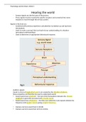
- Sensory signals are the first part of the process.
- These signals must be received by specific receptors and converted into nerve
impulses to travel through the nervous system.
Signals in the brain are:
- Combined with previous experience and attention (so bottom up and top down
information).
- Used to create a percept that can feed into our understanding of a situation
(perceptual understandings)
- Used to determine an appropriate behavioural response.
Auditory signals:
Sound is a form of longitudinal wave in air created by the vibration of objects.
Wave properties can therefore be used to understand sound:
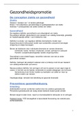
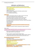
• Sound waves have amplitude – the size of the fluctuations indicates this. Greater
amplitude is generally associated with a louder sound.
• Sound waves have a frequency – the time over which the cycle repeats indicate the
frequency with quicker repeats giving a higher frequency.
• Humans can here sound from 0-140 dB.
• Humans can here sound from 20 Hz to 20 KHz.
, Psychology and the Brain: Week 3
- Volume-left
- Pitch-right
AIR PRESSURE
AIR PRESSURE
TIME TIME
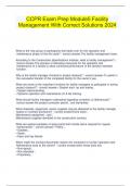
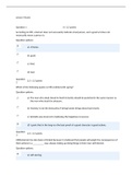
The structure of the ear:
- Pinna
- Auricle- opening of ear
- Extends to external auditory canal
- Tympanic membrane/eardrum
- Malleus, incus, stapes- bones that move in lever-type fashion to transmit
information from eardrum to cochlea (inner ear)
- Semi-circular canals- for balance (vestibular system)
- In cochlea we find two types of hair cells:
- Inner hair cells- detecting auditory stimuli
- Outer hair cells- seems to have role in amplifying signals
- Cochlea nerve and vestibular nerve- combine to form one of the cranial nerves
- Carry information into brain
, Psychology and the Brain: Week 3
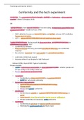
The structure of the ear is highly specialized to support our sense of hearing:
• It can be divided into outer (brown/green), middle (red) and inner (purple).
• The outer ear consists of the part we can easily see and touch: pinna, auricle,
external auditory canal.
• The tympanic membrane separates the outer ear from the air-filled middle ear cavity
which contains three tiny bones (malleus, incus, stapes).
• The stapes contacts the entrance to the cochlea, which is where the auditory hair
cells are, and so sound waves have to get all the way into the inner ear before they
can be sensed.
Outer ear:
- Funnels sound inwards
- Amplifies the sound by acting as a tube for it to echo in (tube resonator)
- Helps sound localisation in the vertical plane (humans) and other planes (moveable
pinna)
- Protection: water-resistant wax, which is antibacterial and antifungal, acidic
environment, hairs prevent entry
Middle ear:
- Protection: middle ear reflex can lock position of bones to prevent transmission of
loud sounds
- Acoustic impedance matching: amplifies pressure created by sound wave to prevent
loss of signal
- Loss of signal due to fluid reflecting sound waves – sound waves can’t pass as
effectively
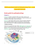
Inner ear:
- Cochlea snail-like shape
- oval window-opening where information transmitted through from middle ear
- information transmitted causes fluid in cochlea to move around snail structure-
forward and back
Hearing the world
- Sensory signals are the first part of the process.
- These signals must be received by specific receptors and converted into nerve
impulses to travel through the nervous system.
Signals in the brain are:
- Combined with previous experience and attention (so bottom up and top down
information).
- Used to create a percept that can feed into our understanding of a situation
(perceptual understandings)
- Used to determine an appropriate behavioural response.
Auditory signals:
Sound is a form of longitudinal wave in air created by the vibration of objects.
Wave properties can therefore be used to understand sound:
• Sound waves have amplitude – the size of the fluctuations indicates this. Greater
amplitude is generally associated with a louder sound.
• Sound waves have a frequency – the time over which the cycle repeats indicate the
frequency with quicker repeats giving a higher frequency.
• Humans can here sound from 0-140 dB.
• Humans can here sound from 20 Hz to 20 KHz.
, Psychology and the Brain: Week 3
- Volume-left
- Pitch-right
AIR PRESSURE
AIR PRESSURE
TIME TIME
The structure of the ear:
- Pinna
- Auricle- opening of ear
- Extends to external auditory canal
- Tympanic membrane/eardrum
- Malleus, incus, stapes- bones that move in lever-type fashion to transmit
information from eardrum to cochlea (inner ear)
- Semi-circular canals- for balance (vestibular system)
- In cochlea we find two types of hair cells:
- Inner hair cells- detecting auditory stimuli
- Outer hair cells- seems to have role in amplifying signals
- Cochlea nerve and vestibular nerve- combine to form one of the cranial nerves
- Carry information into brain
, Psychology and the Brain: Week 3
The structure of the ear is highly specialized to support our sense of hearing:
• It can be divided into outer (brown/green), middle (red) and inner (purple).
• The outer ear consists of the part we can easily see and touch: pinna, auricle,
external auditory canal.
• The tympanic membrane separates the outer ear from the air-filled middle ear cavity
which contains three tiny bones (malleus, incus, stapes).
• The stapes contacts the entrance to the cochlea, which is where the auditory hair
cells are, and so sound waves have to get all the way into the inner ear before they
can be sensed.
Outer ear:
- Funnels sound inwards
- Amplifies the sound by acting as a tube for it to echo in (tube resonator)
- Helps sound localisation in the vertical plane (humans) and other planes (moveable
pinna)
- Protection: water-resistant wax, which is antibacterial and antifungal, acidic
environment, hairs prevent entry
Middle ear:
- Protection: middle ear reflex can lock position of bones to prevent transmission of
loud sounds
- Acoustic impedance matching: amplifies pressure created by sound wave to prevent
loss of signal
- Loss of signal due to fluid reflecting sound waves – sound waves can’t pass as
effectively
Inner ear:
- Cochlea snail-like shape
- oval window-opening where information transmitted through from middle ear
- information transmitted causes fluid in cochlea to move around snail structure-
forward and back