G Protein-Coupled Receptors (GPCRs)
Dr Graham Ladds
Objectives:
• An appreciation of the importance of GPCRs.
• An understanding of GPCR structure and function.
• Be able to demonstrate knowledge of GPCR interacting proteins.
• An understanding of the process leading to GPCR crystal structures.
• Demonstrate knowledge of arrestin-mediated signalling
• An understanding of agonist bias.
• An understand in of the operational model of agonism as is pertains to GPCRs.
• An understanding of allosterism.
• Obtained an appreciation of modern GPCR research.
Essay Titles
1. GPCRs interact with a wide variety of cellular proteins. Discuss with examples how
these interactions influence GPCR signalling outcomes.
2. Allosteric modulation is the future of GPCR drug design – discuss.
3. Describe, with examples the concept of agonist bias. What implications may this
have for drug discovery?
4. GPCRs are classified into different families. Compare and contrast differences
between these families.
5. “Fundamental transition states of GPCRs are critical in determining their signalling
outcome.” Discuss this statement in terms of agonist bias.
,Lecture 1
GPCRs are the largest superfamily of receptors, the human genome is estimated to have at
least 1000 GPCRs, constituting >1% of the human genome, it includes ~400 non-sensory
GPCRs and ~500 sensory GPCRs.
However, only the functions of ~100 GPCRs are known.
GPCR ligands are very diverse, the receptors bind neurotransmitters, neuropeptides, lipids,
odorants, peptides, photons and so on…
They also make up more than 60% of all prescription drugs in the market and sell for >$200
billion a year in total.
They interact with different intracellular effector systems – 2nd messengers, kinases, ion
channels, transcription factors etc. This allows GPCRs to mediate a wide variety of
responses.
GPCRs have a conserved structure, it contains 7 TM domains. The extracellular N terminus
(usually) binds to the ligand. The cytoplasmic loops (especially loop 3 between helices V and
VI) interact with the G protein. Cytoplasmic C-terminal tail is involved in regulating GPCR
activity. Despite a common overall structure, the amino acid sequences of the different
receptors are quite different and it is these differences that determine the specificity of
ligand binding and determine how the different receptors interact with the different G
proteins.
Heterotrimeric G protein couples receptor to target proteins. G protein (guanine nucleotide
binding protein). It contains 3 subunits (Galpha, Gbeta, Ggamma). It acts as a molecular
switch. They are ON when bound to GTP and OFF when bound to GDP. When ligand binds, a
change in the receptor conformation is induced. This triggers release of GDP from the
Galpha subunit and binding of GTP. GDP to GTP nucleotide exchange is induced by the
receptor and promoted by high GTP/GDP ratio. The Galpha-GTP dissociates from
Gbeta/gamma subunits. Galpha-GTP and Gbeta/gamma change the activity of target
proteins. RGS proteins are regulators of G protein signalling, promoting GTPase activity of
Galpha subunit.
There are 3 mammalian GPCR subfamilies (classified according to >20% sequence
homology):
• Family A – rhodopsin-like GPCRs
• Family B – secretin receptor family
• Family C – metabotropic glutamate (mGlu) and GABA(B) receptor family
There are also others but of less pharmacological importance.
There are 16 different Galpha subunits, 5 Gbeta subunits and 12 Ggamma subunits.
Alpha Subunit Members
Gs Gs, Golf
Gi Gi, Go, Gt, Gg, Gz
Gq Gq, G11, G14, G15, G16
G12 G12, G13
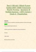
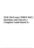
, Signal Integration
Imagine two signaling system (A and B) with a common Effector.
Ligand A binds Receptor A which acts through Mediator A to activate the Effector.
Ligand B binds Receptor B which acts through Mediator B to inhibit the Effector.
The activity of the Effector is determined by the balance between Ligands A and B.
This allows the cell to fine tune its response to take into account more than one signal.
Ligand A Ligand B
Receptor A Receptor B
Stimulate Inhibit
Mediator A Effector Mediator B
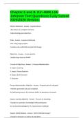
Example of signal integration in cardiomyocytes
In cardiomyocytes, contraction is regulated by stimulatory and inhibitory signals.
Beta-adrenergic receptors stimulate AC while alpha-adrenergic receptors inhibit AC
Both receptors work through G proteins but different alpha subunit families, Gs and Gi,
respectively. Beta-blockers inhibit signaling through the beta-adrenergic receptor. They
reduce the frequency and strength of heart contractions and are used to treat heart failure
by preventing the heart from beating too fast.
-adrenergic receptor -adrenergic receptor
Stimulate Inhibit
Gs protein Adenylate Gi protein
cyclase
Receptor Family 1 – Rhodopsin Family
Examples: dopamine, rhodopsin, vasopressin, oxytocin, cannabinoids, adenosine, serotonin,
opioid receptors, chemokine, somatostatin
Dr Graham Ladds
Objectives:
• An appreciation of the importance of GPCRs.
• An understanding of GPCR structure and function.
• Be able to demonstrate knowledge of GPCR interacting proteins.
• An understanding of the process leading to GPCR crystal structures.
• Demonstrate knowledge of arrestin-mediated signalling
• An understanding of agonist bias.
• An understand in of the operational model of agonism as is pertains to GPCRs.
• An understanding of allosterism.
• Obtained an appreciation of modern GPCR research.
Essay Titles
1. GPCRs interact with a wide variety of cellular proteins. Discuss with examples how
these interactions influence GPCR signalling outcomes.
2. Allosteric modulation is the future of GPCR drug design – discuss.
3. Describe, with examples the concept of agonist bias. What implications may this
have for drug discovery?
4. GPCRs are classified into different families. Compare and contrast differences
between these families.
5. “Fundamental transition states of GPCRs are critical in determining their signalling
outcome.” Discuss this statement in terms of agonist bias.
,Lecture 1
GPCRs are the largest superfamily of receptors, the human genome is estimated to have at
least 1000 GPCRs, constituting >1% of the human genome, it includes ~400 non-sensory
GPCRs and ~500 sensory GPCRs.
However, only the functions of ~100 GPCRs are known.
GPCR ligands are very diverse, the receptors bind neurotransmitters, neuropeptides, lipids,
odorants, peptides, photons and so on…
They also make up more than 60% of all prescription drugs in the market and sell for >$200
billion a year in total.
They interact with different intracellular effector systems – 2nd messengers, kinases, ion
channels, transcription factors etc. This allows GPCRs to mediate a wide variety of
responses.
GPCRs have a conserved structure, it contains 7 TM domains. The extracellular N terminus
(usually) binds to the ligand. The cytoplasmic loops (especially loop 3 between helices V and
VI) interact with the G protein. Cytoplasmic C-terminal tail is involved in regulating GPCR
activity. Despite a common overall structure, the amino acid sequences of the different
receptors are quite different and it is these differences that determine the specificity of
ligand binding and determine how the different receptors interact with the different G
proteins.
Heterotrimeric G protein couples receptor to target proteins. G protein (guanine nucleotide
binding protein). It contains 3 subunits (Galpha, Gbeta, Ggamma). It acts as a molecular
switch. They are ON when bound to GTP and OFF when bound to GDP. When ligand binds, a
change in the receptor conformation is induced. This triggers release of GDP from the
Galpha subunit and binding of GTP. GDP to GTP nucleotide exchange is induced by the
receptor and promoted by high GTP/GDP ratio. The Galpha-GTP dissociates from
Gbeta/gamma subunits. Galpha-GTP and Gbeta/gamma change the activity of target
proteins. RGS proteins are regulators of G protein signalling, promoting GTPase activity of
Galpha subunit.
There are 3 mammalian GPCR subfamilies (classified according to >20% sequence
homology):
• Family A – rhodopsin-like GPCRs
• Family B – secretin receptor family
• Family C – metabotropic glutamate (mGlu) and GABA(B) receptor family
There are also others but of less pharmacological importance.
There are 16 different Galpha subunits, 5 Gbeta subunits and 12 Ggamma subunits.
Alpha Subunit Members
Gs Gs, Golf
Gi Gi, Go, Gt, Gg, Gz
Gq Gq, G11, G14, G15, G16
G12 G12, G13
, Signal Integration
Imagine two signaling system (A and B) with a common Effector.
Ligand A binds Receptor A which acts through Mediator A to activate the Effector.
Ligand B binds Receptor B which acts through Mediator B to inhibit the Effector.
The activity of the Effector is determined by the balance between Ligands A and B.
This allows the cell to fine tune its response to take into account more than one signal.
Ligand A Ligand B
Receptor A Receptor B
Stimulate Inhibit
Mediator A Effector Mediator B
Example of signal integration in cardiomyocytes
In cardiomyocytes, contraction is regulated by stimulatory and inhibitory signals.
Beta-adrenergic receptors stimulate AC while alpha-adrenergic receptors inhibit AC
Both receptors work through G proteins but different alpha subunit families, Gs and Gi,
respectively. Beta-blockers inhibit signaling through the beta-adrenergic receptor. They
reduce the frequency and strength of heart contractions and are used to treat heart failure
by preventing the heart from beating too fast.
-adrenergic receptor -adrenergic receptor
Stimulate Inhibit
Gs protein Adenylate Gi protein
cyclase
Receptor Family 1 – Rhodopsin Family
Examples: dopamine, rhodopsin, vasopressin, oxytocin, cannabinoids, adenosine, serotonin,
opioid receptors, chemokine, somatostatin