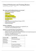
BCHE 5180/6180 Exam Final. 100%
Give the general Henderson-Hasselbalch equation and sketch the plot it describes (pH
against amount of NaOH added to a weak acid). On your curve label the pKa for the
weak acid, and indicate the region in which the buffering capacity of the system is
1 greatest.
8 points
[ A ]
pH pKa log
[HA]
The inflection point, which occurs when
the weak acid has been exactly one half
titrated with NaOH, occurs at a pH equal
to the pKa of the weak acid.
The region of greatest buffering capacity
(where the titration curve is flattest)
occurs at pH values of pKa ±1.
2 Name and briefly define four types of noncovalent interactions that occur between
8 points biological molecules.
(1) Hydrogen bonds: weak electrostatic attractions between one electronegative atom
(such as oxygen or nitrogen) and a hydrogen atom covalently linked to a second
electronegative atom;
(2) electrostatic interactions: relatively weak charge-charge interactions (attractions of
opposite charges, repulsions of like charges) between two ionized groups;
(3) hydrophobic interactions: the forces that tend to bring two hydrophobic groups
together, reducing the total area of the two groups that is exposed to surrounding
molecules of the polar solvent (water);
(4) van der Waals interactions: weak interactions between the electric dipoles that two
close-spaced atoms induce in each other.
, 3 Draw the structure of alanine, leucine, isoleucine, tyrosine, lysine and histidine (At pH
8 points 7.0). Give also the three-letter and the one-letter codes. Indicate the pKa of the side
groups.
(Draw 1 amino acid completely, draw only the R group for the other amino acids)
H
+
H3N C COO-
CH2 OH Tyrosine (Tyr, Y)
CH3 pKa = 10.07
Alanine (Ala, A)
CH3 CH2 CH2 CH2 CH2 NH3+
CH2 CH Leucine (Leu,
CH3 L) Lysine (Lys, K)
pKa = 10.53
CH3 H
CH CH2 CH3 CN
Isoleucine (Ile, I)
CH2 C
CH Histidine (His, H)
N pKa = 6.00
H
4 Explain the differences between common and uncommon amino acids
6 points
The 22 common or coded amino acids are used to make proteins. The proteins are
synthesized on the ribosome where the sequence of amino acids are dictated by the
sequence of codons on an mRNA molecule. The codons are recognized by the anti-
codon on a tRNA molecules that have the amino acid attached. Each coded amino acid
has its own set of tRNA molecules.
Uncommon amino acids include the modified common amino acids as a result of post-
translational modifications, and the group of amino acids that are 1) part of small
polypeptides, 2) are metabolic or 3) are synthetic intermediates.
2