FULL COURSE NOTES BIOC 2580
Henderson Hassle back Equation
- If pH is one unit > pKa, the group is fully deprotonated
- If pH = to pKa, the group is 50% deprotonated and 50% protonated
- If pH is one unit < the pKa, the group is fully protonated
- If pH is less than one unit away from pKa, a calculation needed
- Average N-terminal has pKa of 9.5
- Average C-terminal has pKa of 2.5
- Average side chain has a pKa of 12.5
- Degree of deprotonation
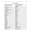
Amino Acid analysis
- Partition /thin layer chromatography (Amino acids exchange between phases, based on polarity)
o Stationary phase: Particles of solid are chosen with a specific property
o Mobile phase: Liquid solvent or buffer flows past the particles and is non polar
o Polar amino acids: spend more of their time hydrogen bonded to silica and move slowly (last)
o Non-polar amino acids: N spend more time in solvent and move fast (first)
o Very polar amino acids have low Rf, non-polar amino acids have high Rf
- Column chromatography
o Sample mix ABC applied at top
o Buffer added carries sample mix through column to collection tubes
o Contents of collection tubes analysed, results plotted
o Volume of buffer needed to move a compound through the column is the elution volume. Compounds
can be identified by their characteristic elution volume
- Ion exchange chromatography
o Separates on the basis of charge, uses charges resins
o Cation exchangers have negative groups which bind positive molecules
o Anion exchangers have positive groups which bind negative molecules
o Elution by changing the pH to alter amino acid so it no longer binds
- How amino acids are detected
o They can be detected by adding ninhydrin which reacts with primary and secondary amine
o Ninhydrin reacts with amino groups to give a purple colour, and is used to detect and measure the
quantity of amino acids on (for example) TLC plates.
o Gives intense purple colour or yellow colour for proline
o Colour intensity is proportional to quantity of amino acid and can be measured
o Alternative is fluoresceine giving yellow fluorescence under UV light
- Metal affinity chromatography
o Clusters of His in a protein bind tightly to Ni or Co
o Column is made up of chelating resin containing Ni
o The His-tagged protein is eluted by adding imidazole to the buffer
o Imidazole out competes His-tag and protein no longer binds to the column
- Separating proteins on the basis of size
o Gel filtration or molecular exclusion chromatography allows separation of proteins on the basis of size
, o Protein molecules can enter the pores if they fit
o Larger proteins are excluded from the pores
- Two dimensional gels
Allow separation of proteins with similar isoelectric points that differ in molecular weight.
- Column chromatography using gel filtration
o Small molecules enter pores and are delayed in their movement down the column
o Intermediate size molecules can only enter some pores and are delayed less
o Very large molecules are excluded from gel and stay in the buffer flowing around the beads… pass
through column quickly
- Electrophoresis
o Separation based on movement of charged molecules in an ELECTRIC FILED
o Positive goes to negative, Negative goes to positive
- SDS PAGE
o the protein is pre-treated with the detergent SDS
o SDS causes protein molecules to extend, and give uniform charge per unit size
o When SDS is used, native protein charge is swamped out and overall negative charge is used to move the
protein through the gel (Towards positive electrode)
o Separation is based on SIZE
o Small proteins fit through the pores and move fast
- Isoelectric focussing
o Separation based on isoelectric point of proteins (the pH a which the net charge on the protein is 0)
o pH where net charge of protein is zero
o At high pH, protein is deprotonated, moves towards the + electrode
o As it passes through gradient of decreasing pH, becomes protonated and negative charge decreases
o Protein stops moving when net charge is 0
- Finding Molar mass of proteins using gel filtration
o Measure elution volume of proteins of known mass
o Elution volume: volume of buffer needed to move a protein from top to bottom of column
o Elution volume is a linear function of log molar mass (negative slope) (x)
o Then measure elution volume of unknown protein and project back to the log mass axis (y)
o Y= mx + b
o Find slope using (Y2-Y1) / (X2 -X1)
Basis of reactivity and hydrolysis
- Peptide bonds of protein are hydrolysed
o Acid hydrolysis
o Base hydrolysis
o With digestive enzymes: proteases
o After hydrolysis, amino acids can be analyzed using chromatography, tells you how much of each amino
acid is present
o Hydrolysis destroys the rest of the polypeptide chain
- Nucleophile: initiate biochemical reactions, nucleus lover, an atom with a lone pair of electrons available to share
o Seek out other groups that are electron deficient (electrophiles)
o Nucleophilic substitution or displacement: incoming nucleophile attacks target atom to displace its
leaving group
o If it has double bond, it only has to lose one of them
o Nucleophilic displacement: Hydrolysis is attack by H2O as nucleophile on the e- deficient C of the peptide
bond
, Determining amino acid sequence
- Sangers method
o can only determine the N-terminal amino acid, ONCE, we can’t repeat it, since hydrolysis destroys the
rest of polypeptide chain
o we can identify the first amino acid in a protein (The N-terminus) by tagging it with fluorodintirobenzene
(bright yellow)
o Flurodinitrobenzene reacts with the amino group of the N-terminal amino acid of a peptide/protein,
which can then be identified after hydrolysis based on the yellow colour of the labelled amino acid.
o The amino acid at the N-terminus of a protein has a free amino group, At high pH, the group
deprotonates to become: NH2-, a nucleophile
o Reacts with fluorodintirobenzene , HF is a good leaving group
- Edman degradation method
o the two step process keeps the reaction cycle in phase so only one amino acid is released per
cycle
o exclusion of H2O prevents hydrolysis of the remaining peptide bonds
o reactions can be repeated
o Step 1: (basic phase) coupling requires base: reaction must be complete before the next cyclization step
can take place (labels the N-terminal AA with PITC)
▪ Important because The N-terminal amino group becomes deprotonated under alkaline
conditions making it nucleophilic and allowing it to react with the electrophilic PITC reagent.
o Step 2: (acidic phase) reaction must be complete before the next coupling step can take place (cleaves
the first peptide bond). Coupling and cyclization cannot occur simutaneously
Selective hydrolysis
- Trypsin
o Cuts peptide bonds to the right of Lys and Arg (L or K)… but not if next to proline
▪ H-bonding groups of trypsin peptide backbone to enzyme , Selected side chain fits in a
long narrow pocket in trypsin, Negative charge at bottom of pocket attracts positive Lys
or Arg. AA that are too large will not fit
- Chymotrypsin
o Cuts peptide bonds to the right of Phe, Tyr and Trp (F, Y or W)… but not if next to proline
▪ Selected side chain fits in a nonpolar pocket in the chymotrypsin, Pocket is large, to fit
aromatic amino acids, benzene ring makes good contact with pocket… small AA may
allow H2O into non polar pocket
o Proximity and orientation: The binding of a large amino acid in the binding pocket of chymotrypsin
positions the peptide bond to be broken close to the catalytic unit.
o General acid/base catalysis: His-57 donates or accepts protons during catalysis.
o Hydrophobic effect: The binding pocket of chymotrypsin is lined with non-polar amino acids.
o Van der Waals forces: The binding pocket of chymotrypsin is the right size to fit a large amino acid.
o Lowering the energy of activation: Chymotrypsin breaks the peptide hydrolysis reaction into two easier
steps.
o Complementary to the transition state: The oxyanion hole binds to the tetrahedral carboxyanion.
- Cyanogen bromide
o Cuts to the right of Met (on the S)
o Peptide chain is broken on carboxylate side and Met is converted to homoserine, Hse
- Overlap method
o Two samples of the original polypeptide are each cut separately using two hydrolysis methods, each
targeting different sites (trypsin, chymotrypsin)
o Sequences from one set of oligopeptides are lined up to overlap with oligopeptides from the other set,
to deduce how they were originally joined
, - Mass Spectrometry
o Cleave one peptide bond per molecule, in random manner
o Flip the order to make it go from N-C
Secondary structure
- regular repetitive patterns such as helix, in short sections of the polypeptide chain
- The polypeptide chain forms a backbone which appears to be linked by C-C and C-N single bonds
- Single bonded structures are flexible due to bond rotation
- Groups connected by single bonds can rotate about bond axis
- Chain flexibility arises from bond rotation not bond bending
- Linus Pauling
o The peptide bond has double bond character
o The peptide bond has two resonance forms, one with a double bond
o Pauling compared lengths of C-N bonds to correlate bond length with bond order
- Alpha helix
o Peptide bonds form rigid planes connecting tetrahedral alpha-C atoms
o Restricted bond rotation limits freedom of motion, so that only a few regular structures can form
o in a helical shape, every a-C bond down the peptide chain turns in same direction (e.g.clockwise)
o in an extended shape, the a-C bonds turn in alternate directions down the peptide chain
o Distance between each helix turn is 5.4 Å
o Number of hydrogen bonds = n -5 +1
o Oxygen on amino 1 will always match up with Hydrogen on amino 5
o Ala, Arg, Gln, Glu, His, Leu, Lys, Met, (Phe) tend to form a-helix
- Beta strand/sheet
o Strands in the SAME direction make a parallel B-sheet
▪ H-bonds connect strand to strand
o Strands in OPPOSITE directions make anti-parallel B-sheet
▪ H-bonds align better in antiparallel mode
o Maximum space available for bulky or awkward shaped side chains
o Distance between each strand is 7 Å
o Trp, Tyr, (Phe), Val, Ile, Thr, Cys need room, prefer b-sheet structure
- Secondary Structure breakers
o GPNDS: Gly, Pro, Asn, Asp ,Ser
o 2 breakers in a group of 4 amino acids interrupts the secondary structure
o Forms a turn or flexible loop
Tertiary structure
- is the overall pattern of folding of the whole polypeptide chain, fiborous proteins
- The simplest possible tertiary structure is continuous secondary structure
- Most proteins are globular: this requires the polypeptide to fold back on itself (causing breaks)
- Allows for flexible loops and turns where polypeptide can change direction to allow folding
- The hydrophobic effect
o is a major force driving protein folding
o Non-polar Aas group together to minimize contact with H2O (hydrophobic effect).. in core
o Polar Aas form outer layer, interact well with surrounding H2O (good H-bonding) or with ions in solution
o Close contacts attract by weak van de Waals forces… Aka London dispersive forces
- A sequence with mostly groups of a-helix-forming AAs will fold into an a-helix bundle
o Non-polar AAs every 3 or 4 places in the helix make a non-polar patch or stripe e.g. -PPNPPNNP-, which
fold to inside of bundle
o AAs that prefer b-sheet are present, but scattered
- B-sheet-forming amino acids in majority fold into antiparallel B-sheet
Henderson Hassle back Equation
- If pH is one unit > pKa, the group is fully deprotonated
- If pH = to pKa, the group is 50% deprotonated and 50% protonated
- If pH is one unit < the pKa, the group is fully protonated
- If pH is less than one unit away from pKa, a calculation needed
- Average N-terminal has pKa of 9.5
- Average C-terminal has pKa of 2.5
- Average side chain has a pKa of 12.5
- Degree of deprotonation
Amino Acid analysis
- Partition /thin layer chromatography (Amino acids exchange between phases, based on polarity)
o Stationary phase: Particles of solid are chosen with a specific property
o Mobile phase: Liquid solvent or buffer flows past the particles and is non polar
o Polar amino acids: spend more of their time hydrogen bonded to silica and move slowly (last)
o Non-polar amino acids: N spend more time in solvent and move fast (first)
o Very polar amino acids have low Rf, non-polar amino acids have high Rf
- Column chromatography
o Sample mix ABC applied at top
o Buffer added carries sample mix through column to collection tubes
o Contents of collection tubes analysed, results plotted
o Volume of buffer needed to move a compound through the column is the elution volume. Compounds
can be identified by their characteristic elution volume
- Ion exchange chromatography
o Separates on the basis of charge, uses charges resins
o Cation exchangers have negative groups which bind positive molecules
o Anion exchangers have positive groups which bind negative molecules
o Elution by changing the pH to alter amino acid so it no longer binds
- How amino acids are detected
o They can be detected by adding ninhydrin which reacts with primary and secondary amine
o Ninhydrin reacts with amino groups to give a purple colour, and is used to detect and measure the
quantity of amino acids on (for example) TLC plates.
o Gives intense purple colour or yellow colour for proline
o Colour intensity is proportional to quantity of amino acid and can be measured
o Alternative is fluoresceine giving yellow fluorescence under UV light
- Metal affinity chromatography
o Clusters of His in a protein bind tightly to Ni or Co
o Column is made up of chelating resin containing Ni
o The His-tagged protein is eluted by adding imidazole to the buffer
o Imidazole out competes His-tag and protein no longer binds to the column
- Separating proteins on the basis of size
o Gel filtration or molecular exclusion chromatography allows separation of proteins on the basis of size
, o Protein molecules can enter the pores if they fit
o Larger proteins are excluded from the pores
- Two dimensional gels
Allow separation of proteins with similar isoelectric points that differ in molecular weight.
- Column chromatography using gel filtration
o Small molecules enter pores and are delayed in their movement down the column
o Intermediate size molecules can only enter some pores and are delayed less
o Very large molecules are excluded from gel and stay in the buffer flowing around the beads… pass
through column quickly
- Electrophoresis
o Separation based on movement of charged molecules in an ELECTRIC FILED
o Positive goes to negative, Negative goes to positive
- SDS PAGE
o the protein is pre-treated with the detergent SDS
o SDS causes protein molecules to extend, and give uniform charge per unit size
o When SDS is used, native protein charge is swamped out and overall negative charge is used to move the
protein through the gel (Towards positive electrode)
o Separation is based on SIZE
o Small proteins fit through the pores and move fast
- Isoelectric focussing
o Separation based on isoelectric point of proteins (the pH a which the net charge on the protein is 0)
o pH where net charge of protein is zero
o At high pH, protein is deprotonated, moves towards the + electrode
o As it passes through gradient of decreasing pH, becomes protonated and negative charge decreases
o Protein stops moving when net charge is 0
- Finding Molar mass of proteins using gel filtration
o Measure elution volume of proteins of known mass
o Elution volume: volume of buffer needed to move a protein from top to bottom of column
o Elution volume is a linear function of log molar mass (negative slope) (x)
o Then measure elution volume of unknown protein and project back to the log mass axis (y)
o Y= mx + b
o Find slope using (Y2-Y1) / (X2 -X1)
Basis of reactivity and hydrolysis
- Peptide bonds of protein are hydrolysed
o Acid hydrolysis
o Base hydrolysis
o With digestive enzymes: proteases
o After hydrolysis, amino acids can be analyzed using chromatography, tells you how much of each amino
acid is present
o Hydrolysis destroys the rest of the polypeptide chain
- Nucleophile: initiate biochemical reactions, nucleus lover, an atom with a lone pair of electrons available to share
o Seek out other groups that are electron deficient (electrophiles)
o Nucleophilic substitution or displacement: incoming nucleophile attacks target atom to displace its
leaving group
o If it has double bond, it only has to lose one of them
o Nucleophilic displacement: Hydrolysis is attack by H2O as nucleophile on the e- deficient C of the peptide
bond
, Determining amino acid sequence
- Sangers method
o can only determine the N-terminal amino acid, ONCE, we can’t repeat it, since hydrolysis destroys the
rest of polypeptide chain
o we can identify the first amino acid in a protein (The N-terminus) by tagging it with fluorodintirobenzene
(bright yellow)
o Flurodinitrobenzene reacts with the amino group of the N-terminal amino acid of a peptide/protein,
which can then be identified after hydrolysis based on the yellow colour of the labelled amino acid.
o The amino acid at the N-terminus of a protein has a free amino group, At high pH, the group
deprotonates to become: NH2-, a nucleophile
o Reacts with fluorodintirobenzene , HF is a good leaving group
- Edman degradation method
o the two step process keeps the reaction cycle in phase so only one amino acid is released per
cycle
o exclusion of H2O prevents hydrolysis of the remaining peptide bonds
o reactions can be repeated
o Step 1: (basic phase) coupling requires base: reaction must be complete before the next cyclization step
can take place (labels the N-terminal AA with PITC)
▪ Important because The N-terminal amino group becomes deprotonated under alkaline
conditions making it nucleophilic and allowing it to react with the electrophilic PITC reagent.
o Step 2: (acidic phase) reaction must be complete before the next coupling step can take place (cleaves
the first peptide bond). Coupling and cyclization cannot occur simutaneously
Selective hydrolysis
- Trypsin
o Cuts peptide bonds to the right of Lys and Arg (L or K)… but not if next to proline
▪ H-bonding groups of trypsin peptide backbone to enzyme , Selected side chain fits in a
long narrow pocket in trypsin, Negative charge at bottom of pocket attracts positive Lys
or Arg. AA that are too large will not fit
- Chymotrypsin
o Cuts peptide bonds to the right of Phe, Tyr and Trp (F, Y or W)… but not if next to proline
▪ Selected side chain fits in a nonpolar pocket in the chymotrypsin, Pocket is large, to fit
aromatic amino acids, benzene ring makes good contact with pocket… small AA may
allow H2O into non polar pocket
o Proximity and orientation: The binding of a large amino acid in the binding pocket of chymotrypsin
positions the peptide bond to be broken close to the catalytic unit.
o General acid/base catalysis: His-57 donates or accepts protons during catalysis.
o Hydrophobic effect: The binding pocket of chymotrypsin is lined with non-polar amino acids.
o Van der Waals forces: The binding pocket of chymotrypsin is the right size to fit a large amino acid.
o Lowering the energy of activation: Chymotrypsin breaks the peptide hydrolysis reaction into two easier
steps.
o Complementary to the transition state: The oxyanion hole binds to the tetrahedral carboxyanion.
- Cyanogen bromide
o Cuts to the right of Met (on the S)
o Peptide chain is broken on carboxylate side and Met is converted to homoserine, Hse
- Overlap method
o Two samples of the original polypeptide are each cut separately using two hydrolysis methods, each
targeting different sites (trypsin, chymotrypsin)
o Sequences from one set of oligopeptides are lined up to overlap with oligopeptides from the other set,
to deduce how they were originally joined
, - Mass Spectrometry
o Cleave one peptide bond per molecule, in random manner
o Flip the order to make it go from N-C
Secondary structure
- regular repetitive patterns such as helix, in short sections of the polypeptide chain
- The polypeptide chain forms a backbone which appears to be linked by C-C and C-N single bonds
- Single bonded structures are flexible due to bond rotation
- Groups connected by single bonds can rotate about bond axis
- Chain flexibility arises from bond rotation not bond bending
- Linus Pauling
o The peptide bond has double bond character
o The peptide bond has two resonance forms, one with a double bond
o Pauling compared lengths of C-N bonds to correlate bond length with bond order
- Alpha helix
o Peptide bonds form rigid planes connecting tetrahedral alpha-C atoms
o Restricted bond rotation limits freedom of motion, so that only a few regular structures can form
o in a helical shape, every a-C bond down the peptide chain turns in same direction (e.g.clockwise)
o in an extended shape, the a-C bonds turn in alternate directions down the peptide chain
o Distance between each helix turn is 5.4 Å
o Number of hydrogen bonds = n -5 +1
o Oxygen on amino 1 will always match up with Hydrogen on amino 5
o Ala, Arg, Gln, Glu, His, Leu, Lys, Met, (Phe) tend to form a-helix
- Beta strand/sheet
o Strands in the SAME direction make a parallel B-sheet
▪ H-bonds connect strand to strand
o Strands in OPPOSITE directions make anti-parallel B-sheet
▪ H-bonds align better in antiparallel mode
o Maximum space available for bulky or awkward shaped side chains
o Distance between each strand is 7 Å
o Trp, Tyr, (Phe), Val, Ile, Thr, Cys need room, prefer b-sheet structure
- Secondary Structure breakers
o GPNDS: Gly, Pro, Asn, Asp ,Ser
o 2 breakers in a group of 4 amino acids interrupts the secondary structure
o Forms a turn or flexible loop
Tertiary structure
- is the overall pattern of folding of the whole polypeptide chain, fiborous proteins
- The simplest possible tertiary structure is continuous secondary structure
- Most proteins are globular: this requires the polypeptide to fold back on itself (causing breaks)
- Allows for flexible loops and turns where polypeptide can change direction to allow folding
- The hydrophobic effect
o is a major force driving protein folding
o Non-polar Aas group together to minimize contact with H2O (hydrophobic effect).. in core
o Polar Aas form outer layer, interact well with surrounding H2O (good H-bonding) or with ions in solution
o Close contacts attract by weak van de Waals forces… Aka London dispersive forces
- A sequence with mostly groups of a-helix-forming AAs will fold into an a-helix bundle
o Non-polar AAs every 3 or 4 places in the helix make a non-polar patch or stripe e.g. -PPNPPNNP-, which
fold to inside of bundle
o AAs that prefer b-sheet are present, but scattered
- B-sheet-forming amino acids in majority fold into antiparallel B-sheet